Download Mendeley
Assembly and phosphoregulatory mechanisms of the budding yeast outer kinetochore KMN complex.
Turner, N.N., Zhang, Z., Yang, J., Muir, K.W., McLaughlin, S.H., Morgan, T., Barford, D.(2026) J Cell Biol 225
- PubMed: 41956986 Search on PubMedSearch on PubMed Central
- DOI: https://doi.org/10.1083/jcb.202506015
- Primary Citation Related Structures:
9S4Q, 9S53, 9S5N - PubMed Abstract:
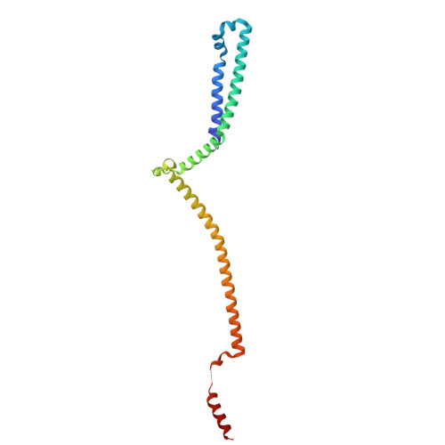
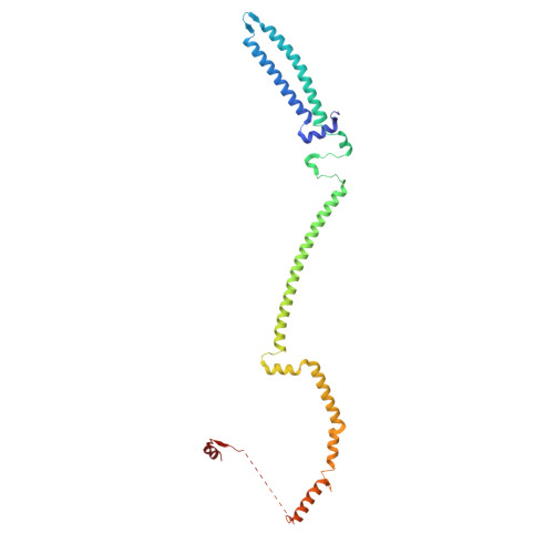
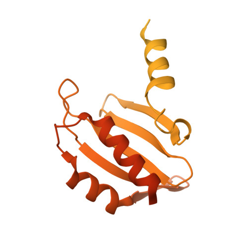
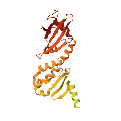
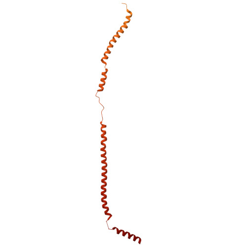
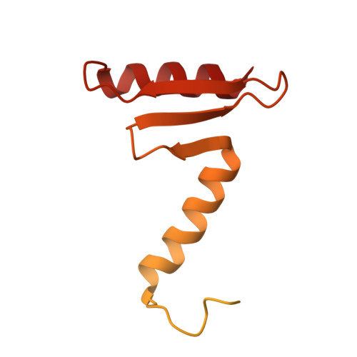
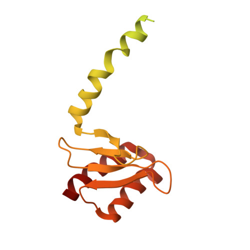
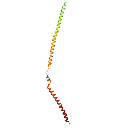
During mitosis and meiosis, kinetochores mediate interactions between chromosomes and spindle microtubules. Kinetochores are multi-megadalton protein complexes essential for chromosome segregation; however, recent structural, functional, and evolutionary studies have revealed divergent mechanisms of kinetochore assembly. Here, we use cryo-EM to understand the structural mechanisms by which the budding yeast microtubule-binding outer kinetochore KMN complex assembles, and how its interactions with the centromere-binding inner kinetochore are regulated. The KMN complex comprises three subcomplexes: Knl1c, Mis12cMtw1c, and Ndc80c. We show how C-terminal motifs of the Mis12cMtw1c subunits Dsn1, Mis12Mtw1, and Nnf1 bind Knl1c and Ndc80c. At the opposite end of the Mis12cMtw1c stalk, an N-terminal auto-inhibitory segment of Dsn1 (Dsn1AI) folds into two α-helices that engage the Mis12cMtw1c head 1 domain, thereby occluding binding sites for the inner kinetochore subunits CENP-CMif2 and CENP-UAme1, reducing their affinity for Mis12cMtw1. Our structure reveals how Aurora BIpl1 phosphorylation of Dsn1AI would release this auto-inhibition to substantially strengthen preexisting connections between the inner and outer kinetochore.
- MRC Laboratory of Molecular Biology , Cambridge, UK.
Organizational Affiliation: