9S1D | pdb_00009s1d

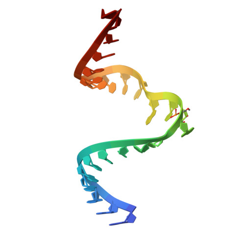
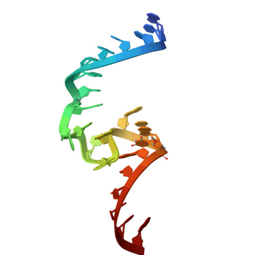
Crystal structure of the methyltransferase ribozyme 1 with two 2'O-methylation (MTR1m2)


Experimental Data Snapshot

  • Method: X-RAY DIFFRACTION
  • Resolution: 2.60 Å
  • R-Value Free: 
    0.251 (Depositor), 0.250 (DCC) 
  • R-Value Work: 
    0.220 (Depositor), 0.219 (DCC) 
  • R-Value Observed: 
    0.222 (Depositor) 

Starting Model: experimental
View more details

wwPDB Validation 3D Report Full Report

Validation slider image for 9S1D

Ligand Structure Quality Assessment 


This is version 1.1 of the entry. See complete history

Literature

A magic methyl effect in the active site of a methyltransferase ribozyme.

Aupic, J.Chen, H.A.Scheitl, C.P.M.Hobartner, C.Magistrato, A.

(2026) Nat Commun 

  • DOI: https://doi.org/10.1038/s41467-026-75575-8
  • Primary Citation Related Structures: 
    9S1D

  • PubMed Abstract: 

    The chemical richness of RNAs is greatly enhanced by post-transcriptional modifications with RNA methylation as the most prominent type. RNA modifications modulate the stability, folding and interaction pattern of RNA molecules. Furthermore, emerging data suggests RNA modifications also directly regulate the activity of catalytic RNA molecules, i.e., ribozymes. Here, we employ classical and hybrid quantum-classical (QM/MM) molecular dynamics (MD) simulations to investigate the reaction mechanism of an artificial methyltransferase ribozyme MTR1. Importantly, we pinpoint how 2'-O-methylations of active site nucleotides synergistically enhance ribozyme activity by reducing the conformational flexibility of the ribose rings and rigidifying the active site. Finally, the herein reported crystal structure of the modified MTR1, solved at 2.6 Å resolution, validates the results of our simulations. Taken together, our work supports the purported central role of modified RNA for early RNA catalysis and may guide rational design of more efficient ribozymes.


  • Organizational Affiliation
    • CNR-Istituto Officina dei Materiali (IOM) c/o Scuola Internazionale Superiore di Studi Avanzati (SISSA), Trieste, Italy. jana.aupic@sissa.it.

Macromolecule Content 

  • Total Structure Weight: 20.11 kDa 
  • Atom Count: 1,374 
  • Modeled Residue Count: 62 
  • Deposited Residue Count: 62 
  • Unique nucleic acid chains: 3

Macromolecules

Find similar nucleic acids by:  Sequence
Entity ID: 1
MoleculeChains LengthOrganismImage
Chains: A14synthetic construct
Sequence Annotations
Expand
Reference Sequence
Find similar nucleic acids by:  Sequence
Entity ID: 2
MoleculeChains LengthOrganismImage
Chains: B24synthetic construct
Sequence Annotations
Expand
Reference Sequence
Find similar nucleic acids by:  Sequence
Entity ID: 3
MoleculeChains LengthOrganismImage
Chains: C24synthetic construct
Sequence Annotations
Expand
Reference Sequence

Experimental Data & Validation

Experimental Data

  • Method: X-RAY DIFFRACTION
  • Resolution: 2.60 Å
  • R-Value Free:  0.251 (Depositor), 0.250 (DCC) 
  • R-Value Work:  0.220 (Depositor), 0.219 (DCC) 
  • R-Value Observed: 0.222 (Depositor) 
Space Group: P 41 21 2
Unit Cell:
Length ( Å )Angle ( ˚ )
a = 71.323α = 90
b = 71.323β = 90
c = 83.512γ = 90
Software Package:
Software NamePurpose
XDSdata reduction
PHASERphasing
Cootmodel building
PHENIXrefinement
XDSdata scaling

Structure Validation

View Full Validation Report


Ligand Structure Quality Assessment 


Entry History 

& Funding Information

Deposition Data


Funding OrganizationLocationGrant Number
German Research Foundation (DFG)Germany--
European Research Council (ERC)European Union--

Revision History  (Full details and data files)

  • Version 1.0: 2026-07-22
    Type: Initial release
  • Version 1.1: 2026-07-29
    Changes: Database references