9S16 | pdb_00009s16

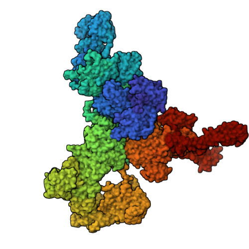
Apo-state RyR1 in the native membrane solved by "SPA


Experimental Data Snapshot

  • Method: ELECTRON MICROSCOPY
  • Resolution: 3.60 Å
  • Aggregation State: PARTICLE 
  • Reconstruction Method: SINGLE PARTICLE 

Starting Model: experimental
View more details

wwPDB Validation 3D Report Full Report

Validation slider image for 9S16

This is version 1.1 of the entry. See complete history. 

Literature

Ligand-induced activation of RyR1 in native membranes.

Mikirtumov, V., Golusik, S., Huo, R., Sprink, T., Balyschew, N., Yang, W., Diebolder, C., Yuan, S., Kotecha, A., Kudryashev, M.

(2026) Nat Commun 17

  • DOI: https://doi.org/10.1038/s41467-026-75504-9
  • Primary Citation Related Structures: 
    9S16, 9S1H, 9S2R, 9S2S, 9S3U, 9S3V, 9S4L, 9S4N, 9S54, 9S55, 9S5A, 9S5C, 9S5E, 9S5F, 9S5W, 9S5Y, 9S5Z

  • PubMed Abstract: 

    Synchronized calcium release through arrays of the ryanodine receptor RyR1, fundamental to skeletal muscle excitation-contraction coupling, is achieved through the mechanical interaction of RyR1s and voltage-sensing receptors DHPR that activate RyR1s in response to action potentials. The calcium release is enhanced through "coupled gating", when the activation of one channel promotes the opening of its neighbours. Here, we determine high-resolution structures of RyR1 in native sarcoplasmic reticulum membranes by cryo-EM/ET, capturing the conformations along the activation pathway and corner-to-corner interfaces between adjacent RyR1 receptors. Compared with purified RyR1s, receptors in native membranes follow an activation pathway with reduced cytosolic-shell tilt and greater consecutive in-plane rotation. Activation-induced rotation remodels the inter-receptor interface, lowering the energy barrier to the cooperative opening of the receptor cluster. Our analysis demonstrates how the native membrane receptor lattice influences ion channel cluster dynamics and provides a mechanistic framework for understanding calcium signaling in muscle.


  • Organizational Affiliation: 
    • In situ Structural Biology, Max Delbrück Center for Molecular Medicine in the Helmholtz Association (MDC), Berlin, Germany.

Macromolecule Content 

  • Total Structure Weight: 2,317.85 kDa 
  • Atom Count: 144,100 
  • Modeled Residue Count: 18,044 
  • Deposited Residue Count: 20,580 
  • Unique protein chains: 2

Macromolecules

Find similar proteins by:|  3D Structure
Entity ID: 1
MoleculeChains  Sequence LengthOrganismDetailsImage
Ryanodine receptor 1
A, B, C, D
5,037Oryctolagus cuniculusMutation(s): 0 
UniProt
Find proteins for P11716 (Oryctolagus cuniculus)
Explore P11716 
Go to UniProtKB:  P11716
Entity Groups
Sequence Clusters30% Identity50% Identity70% Identity90% Identity95% Identity100% Identity
UniProt GroupP11716
Sequence Annotations
Expand
Reference Sequence
Find similar proteins by:|  3D Structure
Entity ID: 2
MoleculeChains  Sequence LengthOrganismDetailsImage
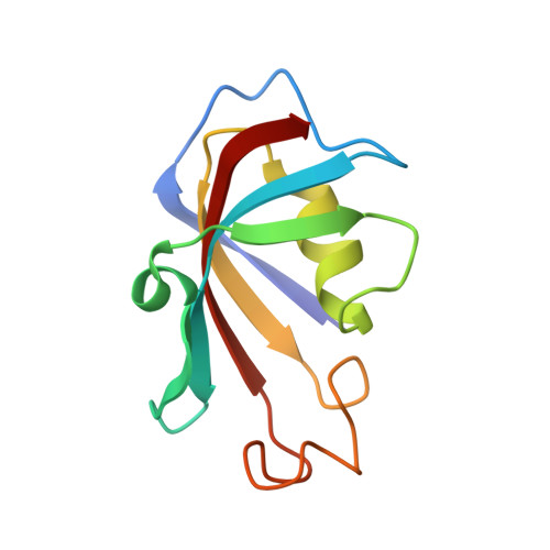
Peptidyl-prolyl cis-trans isomerase FKBP1A
E, F, G, H
108Oryctolagus cuniculusMutation(s): 0 
EC: 5.2.1.8
UniProt
Find proteins for P62943 (Oryctolagus cuniculus)
Explore P62943 
Go to UniProtKB:  P62943
Entity Groups
Sequence Clusters30% Identity50% Identity70% Identity90% Identity95% Identity100% Identity
UniProt GroupP62943
Sequence Annotations
Expand
Reference Sequence

Small Molecules

Ligands 2 Unique
IDChains Name / Formula / InChI Key2D Diagram3D Interactions
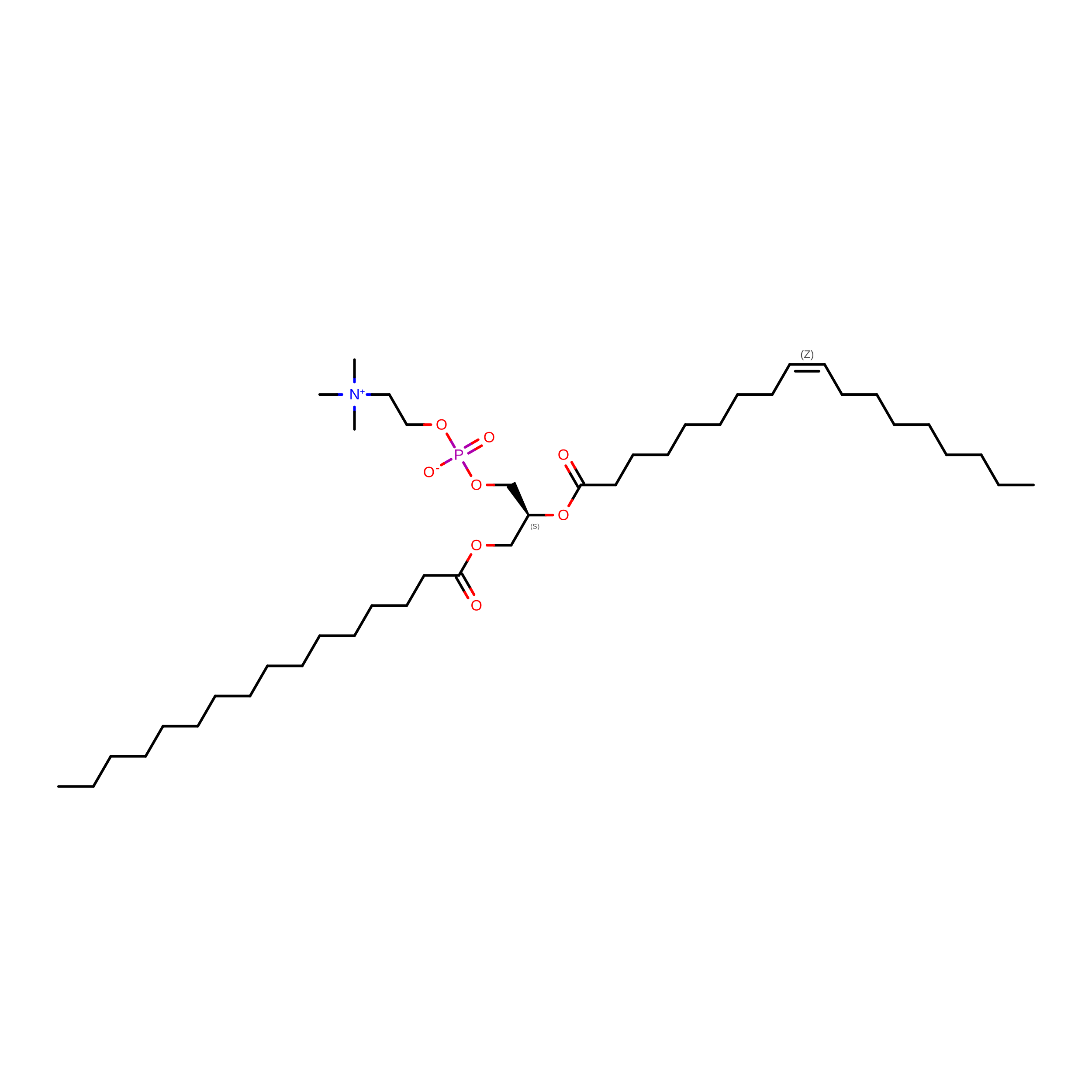
POV
(Subject of Investigation/LOI)

Query on POV


Download:Ideal Coordinates CCD File
I [auth A]
J [auth A]
L [auth B]
M [auth B]
O [auth C]
I [auth A],
J [auth A],
L [auth B],
M [auth B],
O [auth C],
P [auth C],
R [auth D],
S [auth D]
(2S)-3-(hexadecanoyloxy)-2-[(9Z)-octadec-9-enoyloxy]propyl 2-(trimethylammonio)ethyl phosphate
C42 H82 N O8 P
WTJKGGKOPKCXLL-PFDVCBLKSA-N
ZN
(Subject of Investigation/LOI)

Query on ZN


Download:Ideal Coordinates CCD File
K [auth A],
N [auth B],
Q [auth C],
T [auth D]
ZINC ION
Zn
PTFCDOFLOPIGGS-UHFFFAOYSA-N

Experimental Data & Validation

Experimental Data

  • Method: ELECTRON MICROSCOPY
  • Resolution: 3.60 Å
  • Aggregation State: PARTICLE 
  • Reconstruction Method: SINGLE PARTICLE 
EM Software:
TaskSoftware PackageVersion
MODEL REFINEMENTISOLDE
MODEL REFINEMENTPHENIX
RECONSTRUCTIONcryoSPARC4.6.0

Structure Validation

View Full Validation Report


Entry History 

& Funding Information

Deposition Data


Funding OrganizationLocationGrant Number
German Research Foundation (DFG)GermanyKU3221/3-1
Helmholtz AssociationGermany--

Revision History  (Full details and data files)

  • Version 1.0: 2026-07-29
    Type: Initial release
  • Version 1.1: 2026-10-07
    Changes: Data collection, Database references