9RVT | pdb_00009rvt

ACE2 extracellular domain in complex with the macrocyclic peptide GR1.4


Experimental Data Snapshot

  • Method: X-RAY DIFFRACTION
  • Resolution: 2.39 Å
  • R-Value Free: 
    0.298 (Depositor), 0.289 (DCC) 
  • R-Value Work: 
    0.249 (Depositor), 0.246 (DCC) 
  • R-Value Observed: 
    0.250 (Depositor) 

Starting Model: experimental
View more details

wwPDB Validation 3D Report Full Report

Validation slider image for 9RVT

This is version 1.1 of the entry. See complete history

Literature

Yeast Display Technology Enables Rapid Discovery of Low-Nanomolar Macrocyclic Peptide Inhibitors of Human Angiotensin-Converting Enzyme 2.

Romanyuk, Z.Bettin, G.Brear, P.Linciano, S.Mazzocato, Y.Bonadies, S.Zanotto, I.Mazzucco, C.Monferone, A.Soler, M.A.Pasut, G.Martin, S.Scarso, A.Heinis, C.Rothenberger, S.Hyvonen, M.Angelini, A.

(2026) J Med Chem 69: 7689-7708

  • DOI: https://doi.org/10.1021/acs.jmedchem.5c02876
  • Primary Citation Related Structures: 
    28KD, 9RVT, 9SPA

  • PubMed Abstract: 

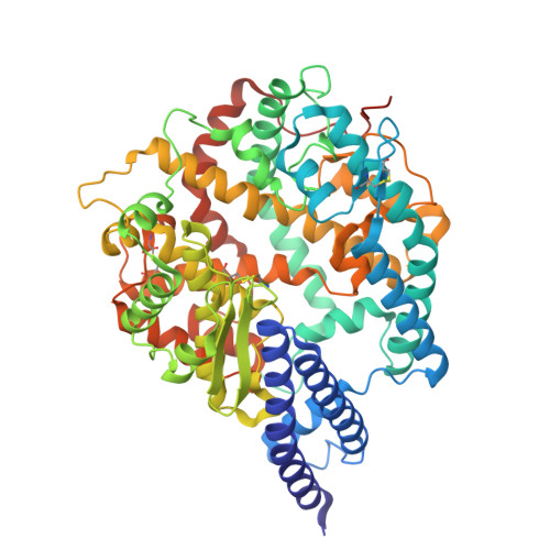
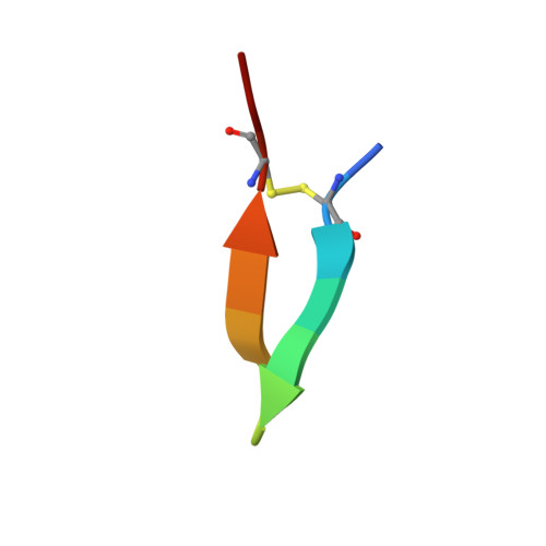
    Macrocyclic peptides (MPs) are valuable molecular formats for drug development, bridging small molecules and larger biologics due to their favorable pharmacological properties. Here, we describe the discovery of low-nanomolar inhibitors of human angiotensin-converting enzyme 2 (hACE2) by quantitatively screening millions of structurally diverse disulfide-cyclized peptide ligands using yeast display technology. The most potent yeast-encoded "one-ring" and "two-ring" MP inhibit hACE2 with K i values of 1.9 and 1.5 nM, respectively. These inhibitory potencies are comparable to those of other cyclic peptides discovered using well-established in vitro display technologies. Crystal structures of the two MPs in complex with hACE2 reveal the adoption of either a rigid β-hairpin or a cysteine-stabilized α-helix/α-helix motif. Both MPs exhibit binding modes distinct from those of previously reported inhibitors. Thus, yeast display is a valid technology to rapidly generate MPs with desired binding properties for the development of potential therapeutics.


  • Organizational Affiliation
    • Department of Molecular Sciences and Nanosystems, Ca' Foscari University of Venice, Via Torino 155, 30172 Mestre, Italy.

Macromolecule Content 

  • Total Structure Weight: 288.3 kDa 
  • Atom Count: 20,476 
  • Modeled Residue Count: 2,438 
  • Deposited Residue Count: 2,492 
  • Unique protein chains: 2

Macromolecules

Find similar proteins by:|  3D Structure
Entity ID: 1
MoleculeChains  Sequence LengthOrganismDetailsImage
Processed angiotensin-converting enzyme 2
A, B, D, G
609Homo sapiensMutation(s): 0 
Gene Names: ACE2UNQ868/PRO1885
EC: 3.4.17 (UniProt), 3.4.17.23 (UniProt)
UniProt & NIH Common Fund Data Resources
Find proteins for Q9BYF1 (Homo sapiens)
Explore Q9BYF1 
Go to UniProtKB:  Q9BYF1
PHAROS:  Q9BYF1
GTEx:  ENSG00000130234 
Entity Groups
Sequence Clusters30% Identity50% Identity70% Identity90% Identity95% Identity100% Identity
UniProt GroupQ9BYF1
Sequence Annotations
Expand
Reference Sequence
Find similar proteins by:  Sequence   |   3D Structure  
Entity ID: 2
MoleculeChains  Sequence LengthOrganismDetailsImage
macrocyclic peptide GR1.4
C, E, F, H
14synthetic constructMutation(s): 0 

Experimental Data & Validation

Experimental Data

  • Method: X-RAY DIFFRACTION
  • Resolution: 2.39 Å
  • R-Value Free:  0.298 (Depositor), 0.289 (DCC) 
  • R-Value Work:  0.249 (Depositor), 0.246 (DCC) 
  • R-Value Observed: 0.250 (Depositor) 
Space Group: P 1 21 1
Unit Cell:
Length ( Å )Angle ( ˚ )
a = 114.234α = 90
b = 77.448β = 101.22
c = 153.688γ = 90
Software Package:
Software NamePurpose
BUSTERrefinement
DIALSdata scaling
XDSdata reduction
PHASERphasing
PDB_EXTRACTdata extraction

Structure Validation

View Full Validation Report


Entry History 

& Funding Information

Deposition Data


Funding OrganizationLocationGrant Number
Other privateUnited Kingdom--

Revision History  (Full details and data files)

  • Version 1.0: 2026-04-08
    Type: Initial release
  • Version 1.1: 2026-04-22
    Changes: Database references