9R3Z | pdb_00009r3z

Exploiting ALDH1A2 and ALDH1A3 Isoform Variability for Crystallization Screening


Experimental Data Snapshot

  • Method: X-RAY DIFFRACTION
  • Resolution: 2.80 Å
  • R-Value Free: 
    0.285 (Depositor), 0.285 (DCC) 
  • R-Value Work: 
    0.208 (Depositor), 0.208 (DCC) 
  • R-Value Observed: 
    0.212 (Depositor) 

Starting Model: experimental
View more details

wwPDB Validation 3D Report Full Report

Validation slider image for 9R3Z

This is version 1.0 of the entry. See complete history

Literature

Exploiting ALDH1A2 and ALDH1A3 isoform variability for crystallisation screening.

Siragusa, S.Garavaglia, S.Mazzorana, M.

(2025) Biochem Biophys Res Commun 780: 152469-152469

  • DOI: https://doi.org/10.1016/j.bbrc.2025.152469
  • Primary Citation Related Structures: 
    9QZE, 9R3Z

  • PubMed Abstract: 

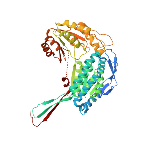
    Members of the human aldehyde dehydrogenase family 1 (ALDH1As) play a crucial role in converting retinal to retinoic acid, a multifunctional bioactive compound. Most evidence highlight ALDH1As as markers for cancer stem cells correlating with tumour aggressiveness. Increasing structural and biochemical data about these important isoenzymes have been reported in literature. Crystal structures of human ALDH1A2 have been so far only obtained in the presence of ligands/cofactors from vapour diffusion hanging drops. Apo-enzyme structures have been described only for the other two members of the family (ALDH1A1 and ALDH1A3) serving as the basis for their co-crystallisation with various ligands. In this study, we describe the first apo-ALDH1A2 structure obtained from nanolitre sitting-drop crystallisation, which expands the potential for high-throughput structure-based drug discovery studies on this isoform. We also explore the crystallisability of NAD + -ALDH1A3 from microlitre drops and compare the structure obtained from it with that of apo-ALDH1A2. Finally, we propose strategies compatible with robotic setups to streamline structural studies on ALDH1A3 and facilitate the exploration of extensive ligand libraries.


  • Organizational Affiliation
    • Department of Scienze del Farmaco, University of Piemonte Orientale, Via Bovio, 6, Novara, 28100, Italy.

Macromolecule Content 

  • Total Structure Weight: 227.13 kDa 
  • Atom Count: 14,710 
  • Modeled Residue Count: 1,892 
  • Deposited Residue Count: 2,072 
  • Unique protein chains: 1

Macromolecules

Find similar proteins by:|  3D Structure
Entity ID: 1
MoleculeChains  Sequence LengthOrganismDetailsImage
Retinal dehydrogenase 2
A, B, C, D
518Homo sapiensMutation(s): 0 
Gene Names: ALDH1A2RALDH2
EC: 1.2.1.36
UniProt & NIH Common Fund Data Resources
Find proteins for O94788 (Homo sapiens)
Explore O94788 
Go to UniProtKB:  O94788
PHAROS:  O94788
GTEx:  ENSG00000128918 
Entity Groups
Sequence Clusters30% Identity50% Identity70% Identity90% Identity95% Identity100% Identity
UniProt GroupO94788
Sequence Annotations
Expand
Reference Sequence

Experimental Data & Validation

Experimental Data

  • Method: X-RAY DIFFRACTION
  • Resolution: 2.80 Å
  • R-Value Free:  0.285 (Depositor), 0.285 (DCC) 
  • R-Value Work:  0.208 (Depositor), 0.208 (DCC) 
  • R-Value Observed: 0.212 (Depositor) 
Space Group: P 1 21 1
Unit Cell:
Length ( Å )Angle ( ˚ )
a = 82.841α = 90
b = 139.478β = 93.927
c = 85.171γ = 90
Software Package:
Software NamePurpose
PHENIXrefinement
xia2data reduction
xia2data scaling
PHASERphasing

Structure Validation

View Full Validation Report


Entry History 

& Funding Information

Deposition Data


Funding OrganizationLocationGrant Number
Italian Ministry of HealthItalyPNRRM6C2-- 12376588

Revision History  (Full details and data files)

  • Version 1.0: 2025-09-03
    Type: Initial release