9QXU | pdb_00009qxu

JetABC DNA loaded state monomer (open)


Experimental Data Snapshot

  • Method: ELECTRON MICROSCOPY
  • Resolution: 3.57 Å
  • Aggregation State: PARTICLE 
  • Reconstruction Method: SINGLE PARTICLE 

Starting Models: experimental, in silico
View more details

wwPDB Validation 3D Report Full Report

Validation slider image for 9QXU

This is version 1.2 of the entry. See complete history

Literature

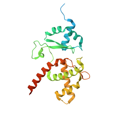
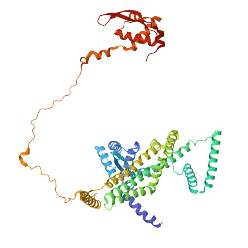
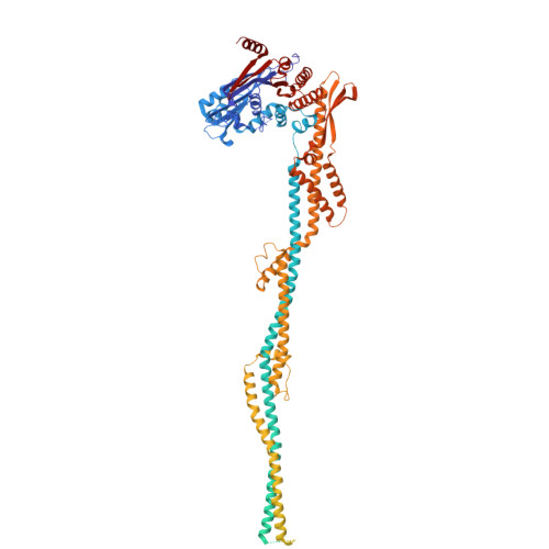
Mechanism of DNA entrapment by a loop-extruding Wadjet SMC motor.

Roisne-Hamelin, F.Liu, H.W.Marechal, N.Uchikawa, E.Durand, A.Gruber, S.

(2025) Mol Cell 85: 3898

  • DOI: https://doi.org/10.1016/j.molcel.2025.09.015
  • Primary Citation Related Structures: 
    9QXR, 9QXS, 9QXT, 9QXU, 9QXV, 9QXX

  • PubMed Abstract: 

    Structural maintenance of chromosome (SMC) complexes perform critical functions by folding DNA through loop extrusion. The choreography and outcome of SMC DNA loading prior to loop extrusion, however, remain elusive. Here, we use cryo-electron microscopy to determine structures of the prokaryotic SMC Wadjet undergoing DNA loading. We show that an initial ATP-triggered relocation of both SMC dimers exposes a DNA-binding pocket and aligns two opened motor units on a DNA double helix. Subsequent ATP hydrolysis drives a nearly 360° rotation of each SMC dimer, closing the motor units around DNA in a sequential manner. This process leads to a DNA-holding conformation-an anticipated key intermediate in loop extrusion-with the DNA held within the kleisin/KITE sub-compartment. Our findings elucidate the mechanism of topological DNA loading by an SMC motor, revealing a straight DNA double helix with motor units oriented tail-to-tail in DNA-holding conformations as the likely starting point of DNA loop extrusion.


  • Organizational Affiliation
    • Department of Fundamental Microbiology (DMF), Faculty of Biology and Medicine (FBM), University of Lausanne (UNIL), 1015 Lausanne, Switzerland.

Macromolecule Content 

  • Total Structure Weight: 458.53 kDa 
  • Atom Count: 23,986 
  • Modeled Residue Count: 2,791 
  • Deposited Residue Count: 3,818 
  • Unique protein chains: 3
  • Unique nucleic acid chains: 1

Macromolecules


Find similar proteins by:|  3D Structure
Entity ID: 1
MoleculeChains  Sequence LengthOrganismDetailsImage
JetBA [auth C],
B [auth D]
250Escherichia coliMutation(s): 0 
Gene Names: ECIG_04711
Find similar proteins by:|  3D Structure
Entity ID: 2
MoleculeChains  Sequence LengthOrganismDetailsImage
JetAC [auth E],
D [auth J]
503Escherichia coliMutation(s): 0 
Gene Names: GP975_00950GQA06_02520
Find similar proteins by:|  3D Structure
Entity ID: 4
MoleculeChains  Sequence LengthOrganismDetailsImage
JetCG [auth A],
H [auth B]
1,096Escherichia coliMutation(s): 0 
Gene Names: C9160_20650
Find similar nucleic acids by:  (by identity cutoff) 
Entity ID: 3
MoleculeChains LengthOrganismImage
Circular plasmid DNA (1843-MER)E [auth P],
F [auth Q]
60Escherichia coli
Sequence Annotations
Expand
Reference Sequence

Experimental Data & Validation

Experimental Data

  • Method: ELECTRON MICROSCOPY
  • Resolution: 3.57 Å
  • Aggregation State: PARTICLE 
  • Reconstruction Method: SINGLE PARTICLE 
EM Software:
TaskSoftware PackageVersion
RECONSTRUCTIONcryoSPARCV4.6
MODEL REFINEMENTPHENIX1.21.2_5419:

Structure Validation

View Full Validation Report


Entry History 

& Funding Information

Deposition Data


Funding OrganizationLocationGrant Number
European Research Council (ERC)European Union724482
Swiss National Science FoundationSwitzerland10001017

Revision History  (Full details and data files)

  • Version 1.0: 2025-10-01
    Type: Initial release
  • Version 1.1: 2025-10-22
    Changes: Data collection, Database references
  • Version 1.2: 2025-11-19
    Changes: Data collection, Database references