9QVR | pdb_00009qvr

Crystal structure of the CtaG_H128A variant from Ruminiclostridium cellulolyticum in complex with PHBA (P2(1)2(1)2(1)-medium)


Experimental Data Snapshot

  • Method: X-RAY DIFFRACTION
  • Resolution: 1.70 Å
  • R-Value Free: 
    0.229 (Depositor), 0.240 (DCC) 
  • R-Value Work: 
    0.203 (Depositor), 0.211 (DCC) 
  • R-Value Observed: 
    0.204 (Depositor) 

Starting Model: experimental
View more details

wwPDB Validation 3D Report Full Report

Validation slider image for 9QVR

Ligand Structure Quality Assessment 


This is version 1.1 of the entry. See complete history

Literature

Distal peptide elongation by a protease-like ligase and two distinct carrier proteins.

Gude, F.Bohne, A.Dell, M.Franke, J.Dunbar, K.L.Groll, M.Hertweck, C.

(2026) Chem 12: None-None

  • DOI: https://doi.org/10.1016/j.chempr.2025.102740
  • Primary Citation Related Structures: 
    9QUZ, 9QVJ, 9QVK, 9QVL, 9QVO, 9QVP, 9QVQ, 9QVR, 9QVS

  • PubMed Abstract: 

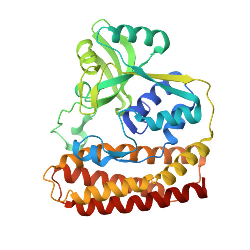
    Closthioamide (CTA) is a potent antibiotic with a unique polythioamide scaffold produced by Ruminiclostridium cellulolyticum . Unlike classical non-ribosomal peptide synthetases (NRPSs), which use modular adenylation and condensation domains, CTA biosynthesis proceeds through non-canonical standalone enzymes. Central to this process is the papain-like ligase CtaG, which catalyzes amide bond formation between two distinct peptidyl carrier proteins (PCPs): CtaH, presenting para-hydroxybenzoic acid (PHBA), and CtaE, carrying a tri-β-alanine ((βAla) 3 ) chain. Using biochemical assays, chemical probes, crystallography, and mutational analysis, we show that CtaG operates via a ping-pong mechanism involving an enzyme-bound intermediate. A single substrate tunnel mediates directional transfer, enabling distal chain elongation that mirrors solid-phase peptide synthesis. Structure-based genome mining revealed homologous enzymes in the biosynthetic pathways of petrobactin, butirosin, and methylolanthanin. Together, our findings uncover a previously overlooked class of thiotemplated ligases and provide a mechanistic blueprint for engineering ribosome-independent peptide assembly lines.


  • Organizational Affiliation
    • Department of Biomolecular Chemistry, Leibniz Institute for Natural Product Research and Infection Biology (Leibniz-HKI), 07743 Jena, Germany.

Macromolecule Content 

  • Total Structure Weight: 74.59 kDa 
  • Atom Count: 5,377 
  • Modeled Residue Count: 612 
  • Deposited Residue Count: 630 
  • Unique protein chains: 1

Macromolecules

Find similar proteins by:|  3D Structure
Entity ID: 1
MoleculeChains  Sequence LengthOrganismDetailsImage
Butirosin biosynthesis protein H N-terminal domain-containing protein
A, B
315Ruminiclostridium cellulolyticumMutation(s): 1 
Gene Names: Ccel_3254
UniProt
Find proteins for B8I0Y7 (Ruminiclostridium cellulolyticum (strain ATCC 35319 / DSM 5812 / JCM 6584 / H10))
Explore B8I0Y7 
Go to UniProtKB:  B8I0Y7
Entity Groups
Sequence Clusters30% Identity50% Identity70% Identity90% Identity95% Identity100% Identity
UniProt GroupB8I0Y7
Sequence Annotations
Expand
Reference Sequence

Experimental Data & Validation

Experimental Data

  • Method: X-RAY DIFFRACTION
  • Resolution: 1.70 Å
  • R-Value Free:  0.229 (Depositor), 0.240 (DCC) 
  • R-Value Work:  0.203 (Depositor), 0.211 (DCC) 
  • R-Value Observed: 0.204 (Depositor) 
Space Group: P 21 21 21
Unit Cell:
Length ( Å )Angle ( ˚ )
a = 59.69α = 90
b = 99.67β = 90
c = 100.74γ = 90
Software Package:
Software NamePurpose
REFMACrefinement
PDB_EXTRACTdata extraction
XDSdata reduction
XSCALEdata scaling
PHASERphasing

Structure Validation

View Full Validation Report


Ligand Structure Quality Assessment 


Entry History 

& Funding Information

Deposition Data


Funding OrganizationLocationGrant Number
German Research Foundation (DFG)Germany--

Revision History  (Full details and data files)

  • Version 1.0: 2025-10-01
    Type: Initial release
  • Version 1.1: 2026-02-18
    Changes: Database references