Download Mendeley
Structural and Biochemical Insights into the Broad-Spectrum TET Enzyme From Methanocaldococcus jannaschii Reveal the Basis of Substrate Specificity in M42 Aminopeptidases.
Atalah, J., Basbous, H., Effantin, G., Kieffer-Jaquinod, S., Schoehn, G., Girard, E., Franzetti, B.(2026) J Mol Biol 438: 169596-169596
- PubMed: 41419167 Search on PubMed
- DOI: https://doi.org/10.1016/j.jmb.2025.169596
- Primary Citation Related Structures:
9QR6 - PubMed Abstract:
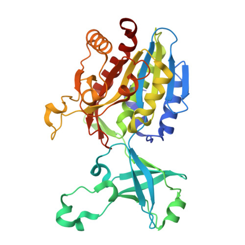
TET peptidases of the M42 family are ∼500 kDa hollow dodecameric complexes ubiquitous in prokaryotes. These enzymes act as strict aminopeptidases, catalyzing the removal of N-terminal amino acids from peptides. A common feature of M42 TET aminopeptidases characterized to date is their marked substrate preference for a limited subset of amino acids. Unlike other hyperthermophilic archaea studied so far, the autotrophic archaeon Methanocaldococcus jannaschii possesses only a single gene encoding an M42 peptidase. This enzyme, named MjTET, is the first reported M42 peptidase to exhibit broad amino acid specificity, including activity on aromatic residues. To assess their peptide degradation efficiencies, the catalytic constants of MjTET were compared to those of its close analogs from Pyrococcus horikoshii. The specialized TETs from P. horikoshii displayed higher catalytic efficiencies than the generalist MjTET, likely reflecting the reliance of Thermococcales on peptide fermentation for energy. Additionally, the structure of MjTET was resolved to 3 Å using cryo-EM and compared with the available models of the four P. horikoshii TETs to identify features underlying substrate specificity. This analysis, combined with mutagenesis studies, revealed a previously uncharacterized loop in the catalytic domain that contributes to substrate discrimination. Collectively, these findings show that substrate specificity in TET enzymes arises from a complex interplay of tertiary structure, oligomeric assembly, and electrostatic surface potential. IMPORTANCE: This study first reported a novel TET peptidase from Methanogenic hyperthermophilic archaea. Its enzymatic properties compared to the specialized TET enzyme characterized so far from heterotrophic archaea suggest a link with autotrophy. It also represents an important step in explaining the structural features guiding substrate specificity.
- Univ Grenoble Alpes, CNRS, CEA, IBS, Grenoble, France.
Organizational Affiliation: