9QJN | pdb_00009qjn

Cryo-EM structure of the human CAK bound to OTS964


Experimental Data Snapshot

  • Method: ELECTRON MICROSCOPY
  • Resolution: 2.40 Å
  • Aggregation State: PARTICLE 
  • Reconstruction Method: SINGLE PARTICLE 

Starting Model: experimental
View more details

wwPDB Validation 3D Report Full Report

Validation slider image for 9QJN

This is version 1.1 of the entry. See complete history

Literature

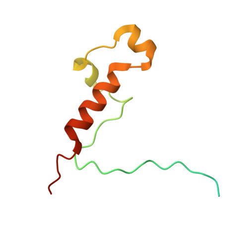
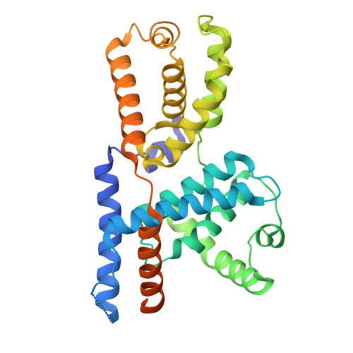
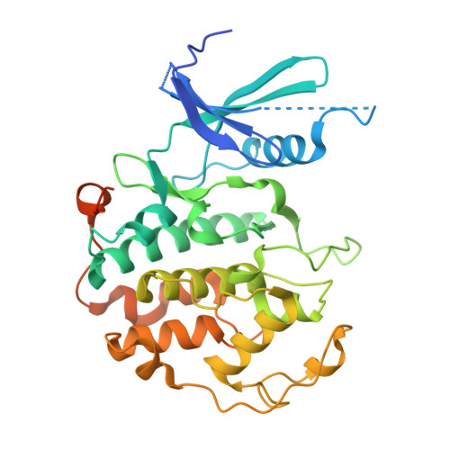
Cryo-EM structures of the CDK11-cyclin L-SAP30BP complex reveal mechanisms of CDK11 regulation.

McGeoch, A.J.S.Cushing, V.I.Roumeliotis, T.I.Cronin, N.B.Hearnshaw, S.J.Choudhary, J.S.Alfieri, C.Greber, B.J.

(2026) Nat Commun 

  • DOI: https://doi.org/10.1038/s41467-026-72329-4

  • PubMed Abstract: 

    The cyclin-dependent kinase CDK11 functions in transcription, mitotic progression, and mRNA splicing. Specifically, spliceosome activation during the B to B act transition depends on phosphorylation of the U2 snRNP component SF3B1 by the CDK11-cyclin L-SAP30BP complex. Here, we present the structure of this spliceosome-activating CDK-cyclin complex, determined by cryogenic electron microscopy at 2.3 Å resolution. Our structure and biochemical experiments show that SAP30BP forms extensive interactions with cyclin L2, thereby stabilising it, and forms critical interactions with the C-terminal kinase lobe of CDK11 that promote complex assembly. Destabilisation of cyclin L2 in the absence of SAP30BP suggests that these principles are applicable to all CDK11-cyclin L complexes. Furthermore, we identify a pseudo-substrate sequence near the CDK11 C-terminus and provide evidence for a role of this segment in CDK11 auto-regulation. Finally, the structure of the CDK11-cyclin L-SAP30BP complex bound to the clinical high-affinity CDK11 inhibitor OTS964 and a comparison to OTS964-bound off-target complexes provide insight into the mechanism of OTS964 selectivity and specificity.


  • Organizational Affiliation
    • Division of Structural Biology, The Institute of Cancer Research, London, UK.

Macromolecule Content 

  • Total Structure Weight: 87.71 kDa 
  • Atom Count: 5,246 
  • Modeled Residue Count: 631 
  • Deposited Residue Count: 766 
  • Unique protein chains: 3

Macromolecules

Find similar proteins by:|  3D Structure
Entity ID: 1
MoleculeChains  Sequence LengthOrganismDetailsImage
CDK-activating kinase assembly factor MAT1A [auth H]93Homo sapiensMutation(s): 0 
Gene Names: MNAT1CAP35MAT1RNF66
UniProt & NIH Common Fund Data Resources
Find proteins for P51948 (Homo sapiens)
Explore P51948 
Go to UniProtKB:  P51948
PHAROS:  P51948
GTEx:  ENSG00000020426 
Entity Groups
Sequence Clusters30% Identity50% Identity70% Identity90% Identity95% Identity100% Identity
UniProt GroupP51948
Sequence Annotations
Expand
Reference Sequence
Find similar proteins by:|  3D Structure
Entity ID: 2
MoleculeChains  Sequence LengthOrganismDetailsImage
Cyclin-HB [auth I]324Homo sapiensMutation(s): 1 
Gene Names: CCNH
UniProt & NIH Common Fund Data Resources
Find proteins for P51946 (Homo sapiens)
Explore P51946 
Go to UniProtKB:  P51946
PHAROS:  P51946
GTEx:  ENSG00000134480 
Entity Groups
Sequence Clusters30% Identity50% Identity70% Identity90% Identity95% Identity100% Identity
UniProt GroupP51946
Sequence Annotations
Expand
Reference Sequence
Find similar proteins by:|  3D Structure
Entity ID: 3
MoleculeChains  Sequence LengthOrganismDetailsImage
Cyclin-dependent kinase 7C [auth J]349Homo sapiensMutation(s): 0 
Gene Names: CDK7CAKCAK1CDKN7MO15STK1
EC: 2.7.11.22 (PDB Primary Data), 2.7.11.23 (PDB Primary Data)
UniProt & NIH Common Fund Data Resources
Find proteins for P50613 (Homo sapiens)
Explore P50613 
Go to UniProtKB:  P50613
PHAROS:  P50613
GTEx:  ENSG00000134058 
Entity Groups
Sequence Clusters30% Identity50% Identity70% Identity90% Identity95% Identity100% Identity
UniProt GroupP50613
Sequence Annotations
Expand
Reference Sequence

Small Molecules

Ligands 1 Unique
IDChains Name / Formula / InChI Key2D Diagram3D Interactions
NK3
(Subject of Investigation/LOI)

Query on NK3


Download:Ideal Coordinates CCD File
D [auth J]OTS964
C23 H24 N2 O2 S
XCFRUAOZMVFDPQ-AWEZNQCLSA-N

Experimental Data & Validation

Experimental Data

  • Method: ELECTRON MICROSCOPY
  • Resolution: 2.40 Å
  • Aggregation State: PARTICLE 
  • Reconstruction Method: SINGLE PARTICLE 
EM Software:
TaskSoftware PackageVersion
MODEL REFINEMENTPHENIX1.21rc1_4985
RECONSTRUCTIONRELION5.0-beta

Structure Validation

View Full Validation Report


Entry History 

& Funding Information

Deposition Data


Funding OrganizationLocationGrant Number
Medical Research Council (MRC, United Kingdom)United KingdomMR/V009354/1

Revision History  (Full details and data files)

  • Version 1.0: 2026-03-18
    Type: Initial release
  • Version 1.1: 2026-05-06
    Changes: Data collection, Database references