Spatial constraints drive amylosome-mediated resistant starch degradation by Ruminococcus bromii in the human colon.
Wimmer, B.H., Morais, S., Amit, I., Tovar-Herrera, O., Tatli, M., Trautwein-Schult, A., Pfister, B., Zalk, R., Todtli, P., Simoni, S., Lisibach, M., Levin, L., Becher, D., Bayer, E.A., Medalia, O., Mizrahi, I.(2025) Nat Commun 16: 10763-10763
- PubMed: 41298524 Search on PubMedSearch on PubMed Central
- DOI: https://doi.org/10.1038/s41467-025-65800-1
- Primary Citation Related Structures:
9QF3, 9QF8, 9QF9, 9QFA - PubMed Abstract:
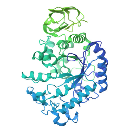
Degradation of complex dietary fiber by gut microbes is essential for colonic fermentation, short-chain fatty acid production, and microbiome function. Ruminococcus bromii is the primary resistant starch (RS) degrader in humans, which relies on the amylosome, a specialized cell-bound enzymatic complex. To unravel its architecture, function, and the interplay among its components, we applied a holistic multilayered approach: Cryo-electron tomography reveals that the amylosome comprises a constitutive extracellular layer extending toward the RS substrate. Proteomics demonstrates remodeling of its contents across different growth conditions, with Amy4 and Amy16 comprising 60% of the amylosome in response to RS. Structural and biochemical analyses reveal complementarity and synergistic RS degradation by these enzymes. We demonstrate that amylosome composition and RS degradation are regulated at two levels: structural constraints and expression-driven shifts in enzyme proportions enforce enzyme proximity, which allows R. bromii to fine-tune its adaptation to dietary fiber and shape colonic metabolism.
- Department of Biochemistry, University of Zurich, Zurich, Switzerland.
Organizational Affiliation: