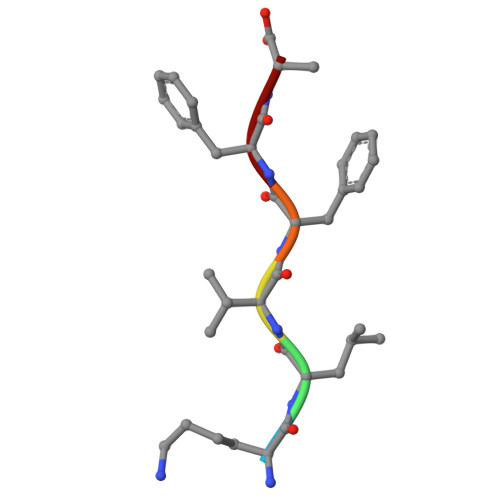
9Q2V | pdb_00009q2v

Structure of peptide segment KLVFFA from amyloid-beta determined by liquid cell MicroED at room temperature

  • Classification: PROTEIN FIBRIL
  • Organism(s): Homo sapiens
  • Mutation(s): No 

  • Deposited: 2025-08-15 Released: 2026-08-26 
  • Deposition Author(s): Vlahakis, N.W., Rodriguez, J.A.
  • Funding Organization(s): Howard Hughes Medical Institute (HHMI), Department of Energy (DOE, United States), National Science Foundation (NSF, United States)

Experimental Data Snapshot

  • Method: ELECTRON CRYSTALLOGRAPHY
  • Resolution: 1.00 Å
  • R-Value Free: 
    0.318 (Depositor), 0.325 (DCC) 
  • R-Value Work: 
    0.276 (Depositor), 0.285 (DCC) 
  • R-Value Observed: 
    0.280 (Depositor) 

Starting Model: experimental
View more details

wwPDB Validation 3D Report Full Report

Validation slider image for 9Q2V

This is version 1.1 of the entry. See complete history. 

Literature

Room Temperature Structures of Solvated Organic Molecules Enabled by Liquid Cell Electron Diffraction.

Vlahakis, N.W., Konieczny, K., Flowers, C.W., Garcia-Garibay, M.A., Rodriguez, J.A.

(2026) ACS Cent Sci 12: 1592-1600

  • DOI: https://doi.org/10.1021/acscentsci.6c00603
  • Primary Citation Related Structures: 
    11ZN, 9Q2V

  • PubMed Abstract: 

    Electron diffraction (MicroED/3DED) enables atomic structure determination from nano- or microcrystals, including dry crystalline powders or mixtures. However, radiation damage and instrument vacuum can limit the interrogation of molecular crystals in solution at room temperature. We now present a strategy for room-temperature MicroED of organic molecular microcrystals suspended in solution, using simple liquid cells: grid sandwiches comprised of two continuous carbon grids that when pressed retain a thin liquid layer between them. In this setting, we find that some room-temperature, hydrated microcrystals are particularly prone to electron beam radiation damage, requiring fast, low dose data collection schemes, direct electron detectors, and multi-crystal data collection. These approaches minimize damage of crystals in liquid cells and facilitate ab initio structures at sub-Ångstrom resolution. This tactic enables study of molecular conformations and crystalline phases that may only be accessible in a room-temperature solvated state.


  • Organizational Affiliation: 
    • Department of Chemistry and Biochemistry, University of California, Los Angeles (UCLA), Los Angeles, California 90095, United States.

Macromolecule Content 

  • Total Structure Weight: 0.78 kDa 
  • Atom Count: 58 
  • Modeled Residue Count: 6 
  • Deposited Residue Count: 6 
  • Unique protein chains: 1

Macromolecules

Find similar proteins by:  Sequence   |   3D Structure  
Entity ID: 1
MoleculeChains  Sequence LengthOrganismDetailsImage
LYS-LEU-VAL-PHE-PHE-ALA6Homo sapiensMutation(s): 0 
Sequence Annotations
Expand
Reference Sequence

Small Molecules

Ligands 1 Unique
IDChains Name / Formula / InChI Key2D Diagram3D Interactions
ACT

Query on ACT


Download:Ideal Coordinates CCD File
B [auth A]ACETATE ION
C2 H3 O2
QTBSBXVTEAMEQO-UHFFFAOYSA-M

Experimental Data & Validation

Experimental Data

  • Method: ELECTRON CRYSTALLOGRAPHY
  • Resolution: 1.00 Å
  • R-Value Free:  0.318 (Depositor), 0.325 (DCC) 
  • R-Value Work:  0.276 (Depositor), 0.285 (DCC) 
  • R-Value Observed: 0.280 (Depositor) 
Space Group: P 21 21 21
Unit Cell:
Length ( Å )Angle ( ˚ )
a = 9.68α = 90
b = 12.94β = 90
c = 41.82γ = 90
EM Software:
TaskSoftware PackageVersion
RECONSTRUCTIONPHENIX

Structure Validation

View Full Validation Report


Entry History 

& Funding Information

Deposition Data


Funding OrganizationLocationGrant Number
Howard Hughes Medical Institute (HHMI)United States--
Department of Energy (DOE, United States)United States--
National Science Foundation (NSF, United States)United States--

Revision History  (Full details and data files)

  • Version 1.0: 2026-08-26
    Type: Initial release
  • Version 1.1: 2026-11-11
    Changes: Database references