Download Mendeley
Overcoming air-water interface-induced artifacts in cryo-EM with protein nanocrates.
Jenkins, M.C., Bobe, D., Johnston, J.D., Cheung, J., Karasawa, A., Zimanyi, C.M., Dermanci, O., McManus, C.T., Finn, M.G., de Marco, A., Kopylov, M.(2026) Nat Methods 23: 1798-1804
- PubMed: 42637978 Search on PubMedSearch on PubMed Central
- DOI: https://doi.org/10.1038/s41592-026-03184-w
- Primary Citation Related Structures:
9Q1B, 9Q1D - PubMed Abstract:
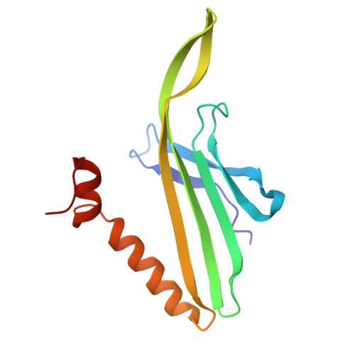
Contact with the air-water interface can bias the orientation of macromolecules during cryo-electron microscopy sample preparation, leading to uneven sample distribution, preferred orientation and damage to the molecules of interest. To prevent this, we describe a method to encapsulate target proteins within highly hydrophilic, structurally homogeneous and stable protein shells, which we refer to as 'nanocrates' for this purpose. Here we describe the packaging, data acquisition and reconstruction of three proof-of-principle examples, each illuminating a different aspect of the method: apoferritin (demonstrating high-resolution), thyroglobulin (solving a known preferred orientation problem) and 7,8-dihydroneopterin aldolase (a structure previously uncharacterized by cryo-electron microscopy).
- School of Chemistry and Biochemistry, School of Biological Sciences, Georgia Institute of Technology, Atlanta, GA, USA.
Organizational Affiliation: