9PRW | pdb_00009prw

Cryo-EM structure of human DNMT3A/3L

  • Classification: TRANSFERASE
  • Organism(s): Homo sapiens
  • Expression System: Escherichia coli BL21(DE3)
  • Mutation(s): No 

  • Deposited: 2025-07-24 Released: 2025-12-31 
  • Deposition Author(s): Lu, J., Song, J.
  • Funding Organization(s): National Institutes of Health/National Institute of General Medical Sciences (NIH/NIGMS)

Experimental Data Snapshot

  • Method: ELECTRON MICROSCOPY
  • Resolution: 3.66 Å
  • Aggregation State: PARTICLE 
  • Reconstruction Method: SINGLE PARTICLE 

wwPDB Validation 3D Report Full Report

Validation slider image for 9PRW

This is version 1.1 of the entry. See complete history

Literature

Structural insight into hierarchical DNMT3A autoinhibition and its dysregulation in disease.

Lu, J.Vig, E.Chen, J.Gretarsson, K.H.Khudaverdyan, N.Shao, Z.Lu, C.Chang, C.A.Song, J.

(2026) Nat Commun 17

  • DOI: https://doi.org/10.1038/s41467-026-69563-1
  • Primary Citation Related Structures: 
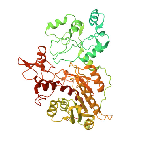
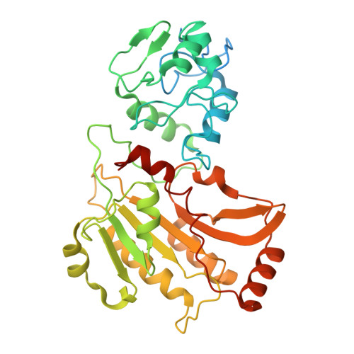
    9PRW

  • PubMed Abstract: 

    DNA methyltransferase DNMT3A-mediated DNA methylation is important for genomic imprinting and transcriptional regulation. However, how the regulatory domains of DNMT3A cooperate with its methyltransferase domain and histone marks to orchestrate genomic methylation remains unclear. Here we report the cryo-EM structure of DNMT3A2 with regulatory factor DNMT3L, revealing an intricate domain interaction underlying multilayered autoinhibition. The PWWP domain interacts with the ADD and methyltransferase domains to block the target recognition domain and the H3K36me2-binding pocket, thereby coupling the H3K36me2 binding with DNMT3A activation, adding a layer of allosteric regulation distinct from the previously characterized ADD-H3K4me0 regulation. Molecular dynamics simulations of the DNMT3A-DNMT3L complex further reveals that relief of DNMT3A autoinhibition involves disengagement of the CpG-recognition loop of the target recognition domain from autoinhibitory interaction, leading to enhanced accessibility of the target recognition domain loop for DNA binding and DNMT3A activation. Importantly, our combined structural, biochemical and genomic methylation analysis demonstrates that disrupting the PWWP-ADD interaction by disease-associated DNMT3A mutations leads to impaired DNMT3A autoinhibition and substrate specificity, providing a potential explanation to aberrant DNA methylation in disease.


  • Organizational Affiliation
    • Department of Biochemistry, University of California, Riverside, CA, USA.

Macromolecule Content 

  • Total Structure Weight: 233.41 kDa 
  • Atom Count: 13,546 
  • Modeled Residue Count: 1,701 
  • Deposited Residue Count: 2,038 
  • Unique protein chains: 2

Macromolecules

Find similar proteins by:|  3D Structure
Entity ID: 1
MoleculeChains  Sequence LengthOrganismDetailsImage
DNA (cytosine-5)-methyltransferase 3A
A, D
633Homo sapiensMutation(s): 0 
Gene Names: DNMT3A
EC: 2.1.1.37 (PDB Primary Data), 2.1.1 (PDB Primary Data)
UniProt & NIH Common Fund Data Resources
Find proteins for Q9Y6K1 (Homo sapiens)
Explore Q9Y6K1 
Go to UniProtKB:  Q9Y6K1
PHAROS:  Q9Y6K1
GTEx:  ENSG00000119772 
Entity Groups
Sequence Clusters30% Identity50% Identity70% Identity90% Identity95% Identity100% Identity
UniProt GroupQ9Y6K1
Sequence Annotations
Expand
Reference Sequence
Find similar proteins by:|  3D Structure
Entity ID: 2
MoleculeChains  Sequence LengthOrganismDetailsImage
DNA (cytosine-5)-methyltransferase 3-like
B, C
386Homo sapiensMutation(s): 0 
Gene Names: DNMT3L
UniProt & NIH Common Fund Data Resources
Find proteins for Q9UJW3 (Homo sapiens)
Explore Q9UJW3 
Go to UniProtKB:  Q9UJW3
PHAROS:  Q9UJW3
GTEx:  ENSG00000142182 
Entity Groups
Sequence Clusters30% Identity50% Identity70% Identity90% Identity95% Identity100% Identity
UniProt GroupQ9UJW3
Sequence Annotations
Expand
Reference Sequence

Small Molecules

Ligands 2 Unique
IDChains Name / Formula / InChI Key2D Diagram3D Interactions
SAH

Query on SAH


Download:Ideal Coordinates CCD File
E [auth A],
O [auth D]
S-ADENOSYL-L-HOMOCYSTEINE
C14 H20 N6 O5 S
ZJUKTBDSGOFHSH-WFMPWKQPSA-N
ZN

Query on ZN


Download:Ideal Coordinates CCD File
F [auth A]
G [auth A]
H [auth A]
I [auth B]
J [auth B]
F [auth A],
G [auth A],
H [auth A],
I [auth B],
J [auth B],
K [auth B],
L [auth C],
M [auth C],
N [auth C],
P [auth D],
Q [auth D],
R [auth D]
ZINC ION
Zn
PTFCDOFLOPIGGS-UHFFFAOYSA-N

Experimental Data & Validation

Experimental Data

  • Method: ELECTRON MICROSCOPY
  • Resolution: 3.66 Å
  • Aggregation State: PARTICLE 
  • Reconstruction Method: SINGLE PARTICLE 
EM Software:
TaskSoftware PackageVersion
MODEL REFINEMENTPHENIX1.21.2_5419:
RECONSTRUCTIONcryoSPARC

Structure Validation

View Full Validation Report


Entry History 

& Funding Information

Deposition Data


Funding OrganizationLocationGrant Number
National Institutes of Health/National Institute of General Medical Sciences (NIH/NIGMS)United States--

Revision History  (Full details and data files)

  • Version 1.0: 2025-12-31
    Type: Initial release
  • Version 1.1: 2026-05-06
    Changes: Data collection, Database references