9PRG | pdb_00009prg

Crystal structure of E.coli DsbA in-complex with analogue 7


Experimental Data Snapshot

  • Method: X-RAY DIFFRACTION
  • Resolution: 1.65 Å
  • R-Value Free: 
    0.218 (Depositor), 0.218 (DCC) 
  • R-Value Work: 
    0.184 (Depositor), 0.184 (DCC) 
  • R-Value Observed: 
    0.186 (Depositor) 

Starting Model: experimental
View more details

wwPDB Validation 3D Report Full Report

Validation slider image for 9PRG

Ligand Structure Quality Assessment 


This is version 1.1 of the entry. See complete history

Literature

Exploiting a Cryptic Pocket in DsbA through Structure-Guided Parallel Synthesis and Direct-to-Biology Screening.

Tasdan, Y.Balaji, G.R.Davidson, J.Akhtar, N.Ilyichova, O.Guetzoyan, L.J.Chandrashekaran, I.R.Alwan, W.Cobb, H.Gunzburg, M.J.Hasanzada, A.Roughley, S.D.Murray, J.B.Thai, V.C.Cliff, T.Kahler, C.M.Mohanty, B.Capuano, B.Doak, B.C.Scanlon, M.J.

(2026) J Med Chem 69: 6760-6774

  • DOI: https://doi.org/10.1021/acs.jmedchem.5c03004
  • Primary Citation Related Structures: 
    9PRE, 9PRF, 9PRG, 9PRH, 9PRI, 9PRJ, 9PRK, 9PRL, 9PRM

  • PubMed Abstract: 

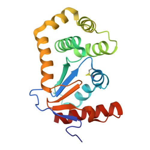
    Antibacterial resistance is a major global health problem, causing an increasing number of deaths worldwide. DsbA, a bacterial oxidoreductase enzyme, is pivotal for the correct folding and activity of virulence factors in bacteria. Inhibiting DsbA presents a promising avenue for developing antivirulence compounds and combating bacterial resistance. The enzyme's structure features two ligand-binding sites: a hydrophobic groove that is the binding site for natural peptide substrates and a "cryptic pocket" enclosed within the protein, which has recently been identified as a target for ligand design. In this study, we report the elaboration of a fragment from within the enclosed cryptic pocket into the hydrophobic groove of Escherichia coli DsbA, using X-ray crystallography-guided structure-based design and parallel synthesis coupled with crude reaction mixture screening (direct-to-biology). This effort yielded the most potent small-molecule Ec DsbA inhibitors reported to date and exemplifies a productive strategy for exploiting a cryptic pocket for drug development.


  • Organizational Affiliation
    • Medicinal Chemistry, Monash Institute of Pharmaceutical Sciences, Monash University, 381 Royal Parade, Parkville, VIC 3052, Australia.

Macromolecule Content 

  • Total Structure Weight: 43.51 kDa 
  • Atom Count: 3,358 
  • Modeled Residue Count: 372 
  • Deposited Residue Count: 376 
  • Unique protein chains: 1

Macromolecules

Find similar proteins by:|  3D Structure
Entity ID: 1
MoleculeChains  Sequence LengthOrganismDetailsImage
Thiol:disulfide interchange protein DsbA
A, B
188Escherichia coliMutation(s): 0 
Gene Names: dsbAdsfppfAb3860JW3832
UniProt
Find proteins for P0AEG4 (Escherichia coli (strain K12))
Explore P0AEG4 
Go to UniProtKB:  P0AEG4
Entity Groups
Sequence Clusters30% Identity50% Identity70% Identity90% Identity95% Identity100% Identity
UniProt GroupP0AEG4
Sequence Annotations
Expand
Reference Sequence

Experimental Data & Validation

Experimental Data

  • Method: X-RAY DIFFRACTION
  • Resolution: 1.65 Å
  • R-Value Free:  0.218 (Depositor), 0.218 (DCC) 
  • R-Value Work:  0.184 (Depositor), 0.184 (DCC) 
  • R-Value Observed: 0.186 (Depositor) 
Space Group: P 1 21 1
Unit Cell:
Length ( Å )Angle ( ˚ )
a = 41.809α = 90
b = 54.515β = 100.717
c = 75.984γ = 90
Software Package:
Software NamePurpose
PHENIXrefinement
Aimlessdata scaling
XDSdata reduction
PHASERphasing
PDB_EXTRACTdata extraction

Structure Validation

View Full Validation Report


Ligand Structure Quality Assessment 


Entry History 

& Funding Information

Deposition Data


Funding OrganizationLocationGrant Number
National Health and Medical Research Council (NHMRC, Australia)Australia--

Revision History  (Full details and data files)

  • Version 1.0: 2026-03-25
    Type: Initial release
  • Version 1.1: 2026-04-08
    Changes: Database references