9PIB | pdb_00009pib

Crystal structure of the A/Puerto Rico/8/1934 (H1N1) influenza virus hemagglutinin in complex with fusion inhibitor CLIPS peptide CP121132 (CP-S1)


Experimental Data Snapshot

  • Method: X-RAY DIFFRACTION
  • Resolution: 2.35 Å
  • R-Value Free: 
    0.231 (Depositor), 0.231 (DCC) 
  • R-Value Work: 
    0.189 (Depositor), 0.190 (DCC) 
  • R-Value Observed: 
    0.191 (Depositor) 

Starting Model: experimental
View more details

wwPDB Validation 3D Report Full Report

Validation slider image for 9PIB

Ligand Structure Quality Assessment 


This is version 1.1 of the entry. See complete history

Literature

Small molecule-constrained paratope mimetic bicyclic peptides as potent inhibitors of group 1 and 2 influenza A virus hemagglutinins.

Kadam, R.U.Juraszek, J.Brandenburg, B.Garg, D.Zhu, X.Jongeneelen, M.Tang, C.Schepens, W.B.G.Buyck, C.Stoops, B.Vermond, J.Vogels, R.Friesen, R.H.E.van Dongen, M.J.P.Wilson, I.A.

(2026) Proc Natl Acad Sci U S A 123: e2537533123-e2537533123

  • DOI: https://doi.org/10.1073/pnas.2537533123
  • Primary Citation Related Structures: 
    9PIB

  • PubMed Abstract: 

    Influenza continues to be a major threat to global health and a substantial economic burden. Innovative strategies are needed to tackle the growing resistance to established influenza therapeutics and to develop new therapeutics with novel mechanisms of action. Previous peptide and small molecule designs have been successful only against influenza group 1 hemagglutinin (HA). Here, we report on a CLIPS (Chemical Linkage of Peptides onto Scaffolds)-based approach to design potent peptidic inhibitors of influenza A viruses that now extend to both group 1 and group 2 HAs. This approach merges features of antibodies and small molecules to design constrained bicyclic peptides that engage the highly conserved HA stem. The heavy-chain complementarity-determining region 3 (HCDR3) of human broadly neutralizing antibody FI6v3 was grafted onto functionalized small molecule scaffolds. The designed peptides exhibited in vitro heterosubtypic cross-reactivity in binding to group 1 (H1 and H5) and group 2 (H3 and H7) HAs and in neutralization of H1N1, H5N1, and H7N3 viruses. A crystal structure of the bicyclic peptide with HA from H1N1 A/Puerto Rico/8/1934 (H1/PR8) at 2.35 Å resolution revealed that the designed peptide faithfully mimics the binding mode and functionality of the parent antibody FI6v3 to the highly conserved stem epitope. These structural and functional data illustrate how both group 1 and group 2 influenza A viruses can now be targeted by constrained peptidic ligands that should aid in development of pan-influenza therapeutics.


  • Organizational Affiliation
    • Department of Integrative Structural and Computational Biology, The Scripps Research Institute, La Jolla, CA 92037.

Macromolecule Content 

  • Total Structure Weight: 238.76 kDa 
  • Atom Count: 17,374 
  • Modeled Residue Count: 2,024 
  • Deposited Residue Count: 2,060 
  • Unique protein chains: 3

Macromolecules

Find similar proteins by:|  3D Structure
Entity ID: 1
MoleculeChains  Sequence LengthOrganismDetailsImage
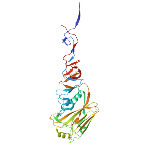
Hemagglutinin
A, C, E, G
326Influenza A virus (A/Puerto Rico/8/1934(H1N1))Mutation(s): 0 
Gene Names: HA
UniProt
Find proteins for P03452 (Influenza A virus (strain A/Puerto Rico/8/1934 H1N1))
Explore P03452 
Go to UniProtKB:  P03452
Entity Groups
Sequence Clusters30% Identity50% Identity70% Identity90% Identity95% Identity100% Identity
UniProt GroupP03452
Glycosylation
Glycosylation Sites: 3
Sequence Annotations
Expand
Reference Sequence
Find similar proteins by:|  3D Structure
Entity ID: 2
MoleculeChains  Sequence LengthOrganismDetailsImage
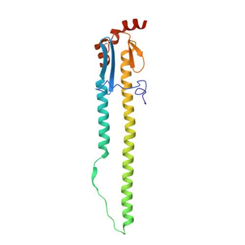
Hemagglutinin HA2 chain
B, D, F, H
176Influenza A virus (A/Puerto Rico/8/1934(H1N1))Mutation(s): 0 
Gene Names: HA
UniProt
Find proteins for P03452 (Influenza A virus (strain A/Puerto Rico/8/1934 H1N1))
Explore P03452 
Go to UniProtKB:  P03452
Entity Groups
Sequence Clusters30% Identity50% Identity70% Identity90% Identity95% Identity100% Identity
UniProt GroupP03452
Glycosylation
Glycosylation Sites: 1
Sequence Annotations
Expand
Reference Sequence
Find similar proteins by:  Sequence   |   3D Structure  
Entity ID: 3
MoleculeChains  Sequence LengthOrganismDetailsImage
CLIPS peptide CP121132I [auth N],
J [auth O],
K [auth P],
L [auth Q]
13synthetic constructMutation(s): 0 

Oligosaccharides

Help  
Entity ID: 4
MoleculeChains Length2D Diagram GlycosylationD Interactions
2-acetamido-2-deoxy-beta-D-glucopyranose-(1-4)-[alpha-L-fucopyranose-(1-6)]2-acetamido-2-deoxy-beta-D-glucopyranoseM [auth J]3N-Glycosylation
Glycosylation Resources
GlyTouCan: G21290RB
GlyCosmos: G21290RB
GlyGen: G21290RB
Entity ID: 5
MoleculeChains Length2D Diagram GlycosylationD Interactions
alpha-L-fucopyranose-(1-3)-[2-acetamido-2-deoxy-beta-D-glucopyranose-(1-4)]2-acetamido-2-deoxy-beta-D-glucopyranoseN [auth I],
P [auth L]
3N-Glycosylation
Glycosylation Resources
GlyTouCan: G11971MR
GlyCosmos: G11971MR
GlyGen: G11971MR
Entity ID: 6
MoleculeChains Length2D Diagram GlycosylationD Interactions
2-acetamido-2-deoxy-beta-D-glucopyranose-(1-4)-2-acetamido-2-deoxy-beta-D-glucopyranoseO [auth K],
Q [auth M]
2N-Glycosylation
Glycosylation Resources
GlyTouCan: G42666HT
GlyCosmos: G42666HT
GlyGen: G42666HT

Small Molecules

Ligands 4 Unique
IDChains Name / Formula / InChI Key2D Diagram3D Interactions
NAG

Query on NAG


Download:Ideal Coordinates CCD File
BA [auth F]
DA [auth G]
GA [auth H]
R [auth A]
V [auth C]
BA [auth F],
DA [auth G],
GA [auth H],
R [auth A],
V [auth C],
X [auth D],
Y [auth E]
2-acetamido-2-deoxy-beta-D-glucopyranose
C8 H15 N O6
OVRNDRQMDRJTHS-FMDGEEDCSA-N
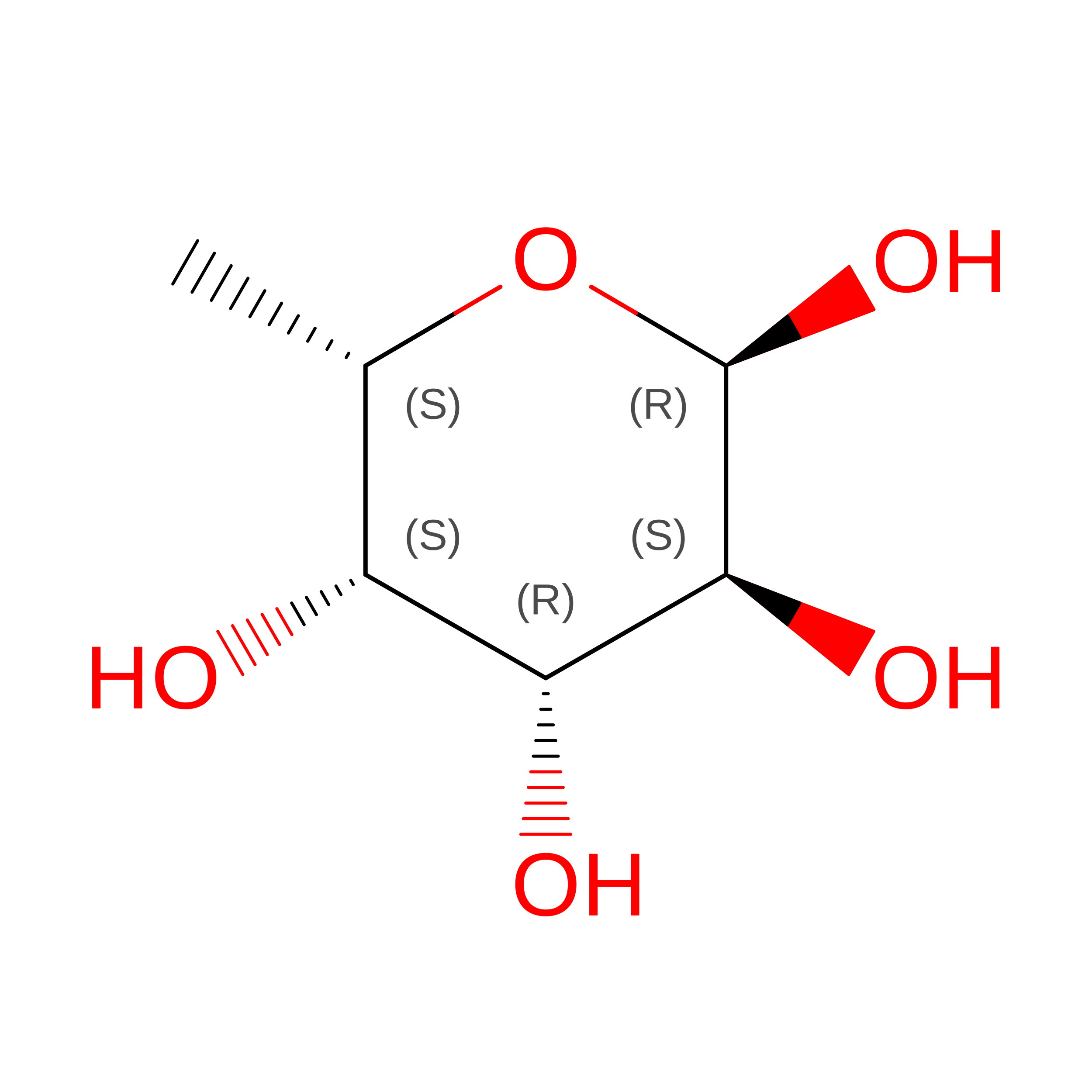
FUC

Query on FUC


Download:Ideal Coordinates CCD File
EA [auth G]alpha-L-fucopyranose
C6 H12 O5
SHZGCJCMOBCMKK-SXUWKVJYSA-N
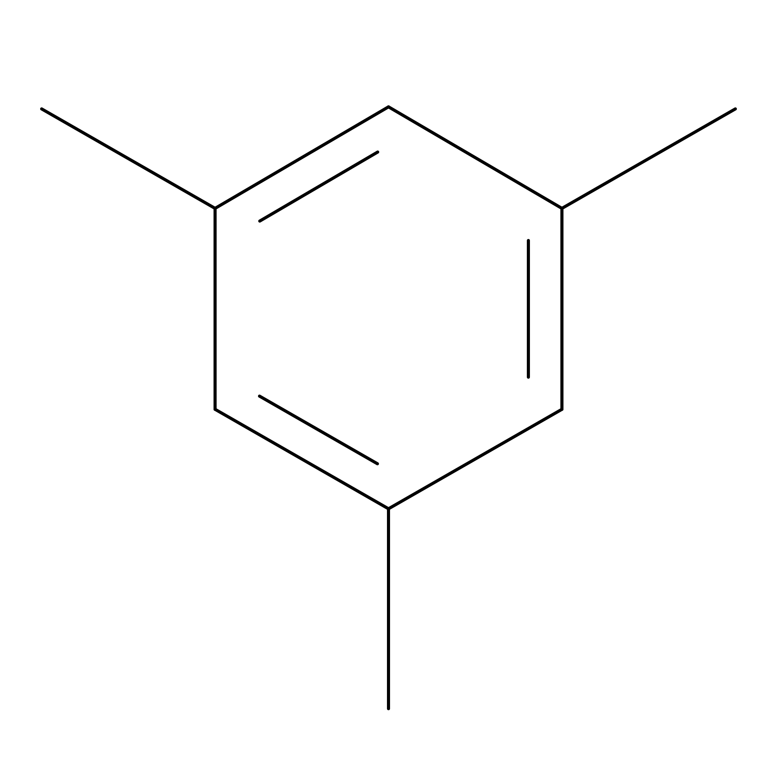
SEZ

Query on SEZ


Download:Ideal Coordinates CCD File
HA [auth N],
KA [auth O],
LA [auth P],
MA [auth Q]
1,3,5-trimethylbenzene
C9 H12
AUHZEENZYGFFBQ-UHFFFAOYSA-N
PEG

Query on PEG


Download:Ideal Coordinates CCD File
AA [auth E]
CA [auth F]
FA [auth G]
IA [auth N]
JA [auth N]
AA [auth E],
CA [auth F],
FA [auth G],
IA [auth N],
JA [auth N],
S [auth A],
T [auth A],
U [auth B],
W [auth C],
Z [auth E]
DI(HYDROXYETHYL)ETHER
C4 H10 O3
MTHSVFCYNBDYFN-UHFFFAOYSA-N

Experimental Data & Validation

Experimental Data

  • Method: X-RAY DIFFRACTION
  • Resolution: 2.35 Å
  • R-Value Free:  0.231 (Depositor), 0.231 (DCC) 
  • R-Value Work:  0.189 (Depositor), 0.190 (DCC) 
  • R-Value Observed: 0.191 (Depositor) 
Space Group: P 3
Unit Cell:
Length ( Å )Angle ( ˚ )
a = 93.623α = 90
b = 93.623β = 90
c = 279.479γ = 120
Software Package:
Software NamePurpose
PHENIXrefinement
PHENIXrefinement
HKL-2000data reduction
HKL-2000data scaling
PHASERphasing

Structure Validation

View Full Validation Report


Ligand Structure Quality Assessment 


Entry History 

& Funding Information

Deposition Data


Funding OrganizationLocationGrant Number
National Institutes of Health/National Institute Of Allergy and Infectious Diseases (NIH/NIAID)United States--

Revision History  (Full details and data files)

  • Version 1.0: 2026-03-18
    Type: Initial release
  • Version 1.1: 2026-04-08
    Changes: Database references