9PI9 | pdb_00009pi9

Sacituzumab Fab bound to Trop2 Dimer


Experimental Data Snapshot

  • Method: X-RAY DIFFRACTION
  • Resolution: 1.56 Å
  • R-Value Free: 
    0.211 (Depositor), 0.210 (DCC) 
  • R-Value Work: 
    0.181 (Depositor), 0.180 (DCC) 
  • R-Value Observed: 
    0.181 (Depositor) 

Starting Model: experimental
View more details

wwPDB Validation 3D Report Full Report

Validation slider image for 9PI9

Ligand Structure Quality Assessment 


This is version 1.2 of the entry. See complete history

Literature

The therapeutic antibody sacituzumab induces trophoblast cell-surface antigen-2 conformational rearrangement.

Ferrao, R.Zhang, J.Chou, C.C.Nieto, A.Langeslay, D.Chatterjee, M.Jin, D.Hung, M.Scott, I.Nagel, M.Xing, W.Letarte, S.Wang, J.Ambrogelly, A.Lansdon, E.B.

(2026) Structure 34: 264

  • DOI: https://doi.org/10.1016/j.str.2025.11.002
  • Primary Citation Related Structures: 
    9PI9

  • PubMed Abstract: 

    Sacituzumab govitecan (SG) is a therapeutic antibody-drug conjugate globally approved for the treatment of breast cancer. SG targets the trophoblast cell-surface antigen-2 (Trop2) at the surface of cancer cells to deliver the cytotoxic topoisomerase I inhibitor SN-38 to the tumor microenvironment. SN-38 is covalently linked to the humanized monoclonal antibody (mAb) sacituzumab via a hydrolyzable linker. Here, we describe the 1.56-Å X-ray crystal structure and stoichiometry of the human Trop2 ectodomain in complex with a sacituzumab (hRS7) antigen-binding Fab fragment. The complex reveals a 2:2 stoichiometry where two sacituzumab Fabs bind across the two Trop2 dimer subunits, inducing a conformational change compared to the apo-structure. Cryo-electron microscopy (cryoEM) and size-exclusion chromatography in combination with multi-angle light scattering (SEC-MALS) analysis of the intact sacituzumab mAb bound to the Trop2 ECD reveals a complex whereby sacituzumab engages two Trop2 dimers in a 2:4 stoichiometry.


  • Organizational Affiliation
    • Gilead Sciences Inc., Structural Biology and Chemistry, 333 Lakeside Dr., Foster City, CA 94404, USA.

Macromolecule Content 

  • Total Structure Weight: 153.73 kDa 
  • Atom Count: 11,459 
  • Modeled Residue Count: 1,315 
  • Deposited Residue Count: 1,380 
  • Unique protein chains: 3

Macromolecules

Find similar proteins by:|  3D Structure
Entity ID: 1
MoleculeChains  Sequence LengthOrganismDetailsImage
Sacituzumab Heavy Chain
A, C
226Homo sapiensMutation(s): 0 
Find similar proteins by:|  3D Structure
Entity ID: 2
MoleculeChains  Sequence LengthOrganismDetailsImage
Sacituzumab Light Chain
B, D
214Homo sapiensMutation(s): 0 
Find similar proteins by:|  3D Structure
Entity ID: 3
MoleculeChains  Sequence LengthOrganismDetailsImage
Tumor-associated calcium signal transducer 2
E, F
250Homo sapiensMutation(s): 0 
Gene Names: TACSTD2GA733-1M1S1TROP2
UniProt & NIH Common Fund Data Resources
Find proteins for P09758 (Homo sapiens)
Explore P09758 
Go to UniProtKB:  P09758
PHAROS:  P09758
GTEx:  ENSG00000184292 
Entity Groups
Sequence Clusters30% Identity50% Identity70% Identity90% Identity95% Identity100% Identity
UniProt GroupP09758
Glycosylation
Glycosylation Sites: 1Go to GlyGen: P09758-1
Sequence Annotations
Expand
Reference Sequence

Oligosaccharides

Help  
Entity ID: 4
MoleculeChains Length2D Diagram GlycosylationD Interactions
2-acetamido-2-deoxy-beta-D-glucopyranose-(1-4)-2-acetamido-2-deoxy-beta-D-glucopyranose
G
2N-Glycosylation
Glycosylation Resources
GlyTouCan: G42666HT
GlyCosmos: G42666HT
GlyGen: G42666HT

Small Molecules

Ligands 4 Unique
IDChains Name / Formula / InChI Key2D Diagram3D Interactions
NAG

Query on NAG


Download:Ideal Coordinates CCD File
T [auth E]2-acetamido-2-deoxy-beta-D-glucopyranose
C8 H15 N O6
OVRNDRQMDRJTHS-FMDGEEDCSA-N
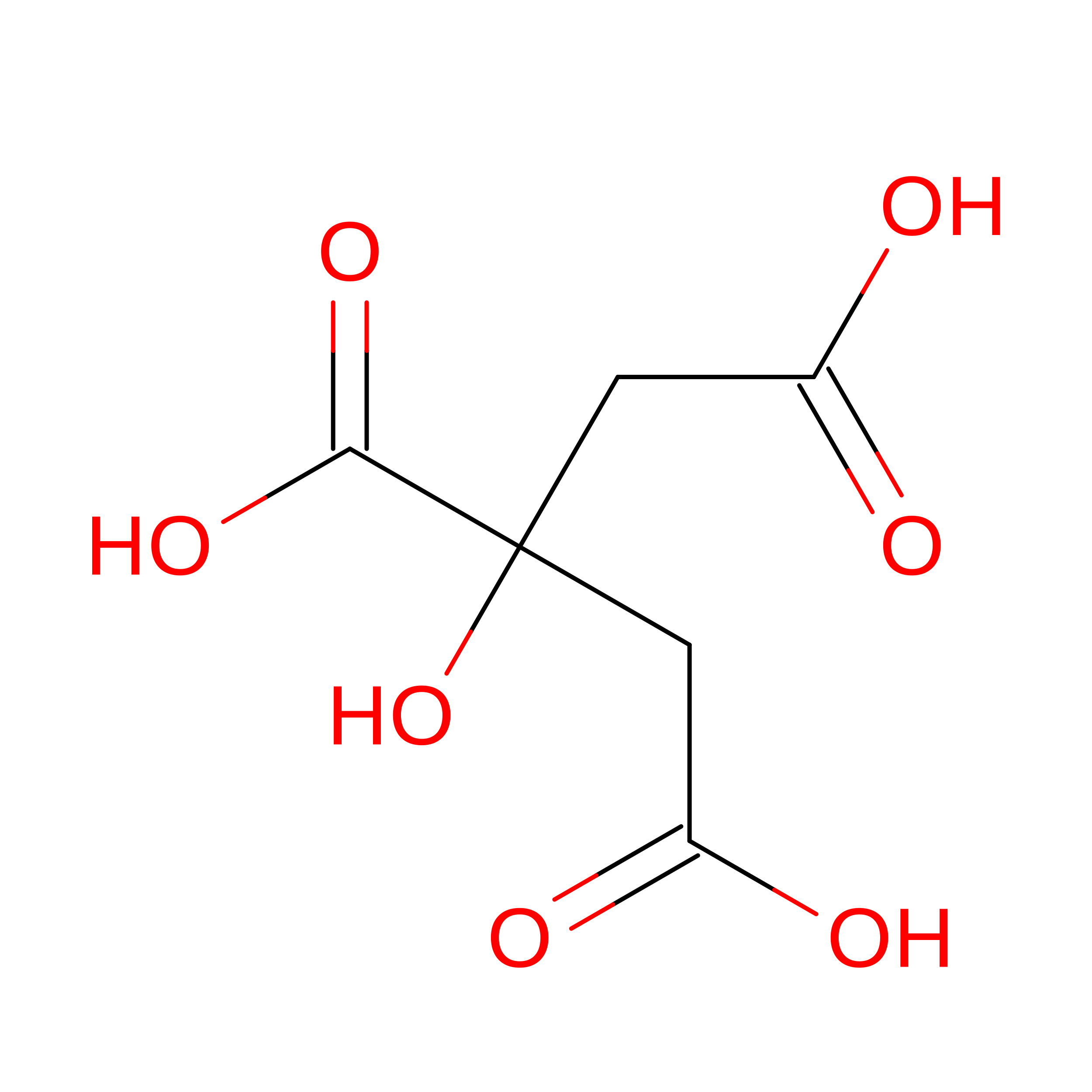
CIT

Query on CIT


Download:Ideal Coordinates CCD File
U [auth E],
W [auth F]
CITRIC ACID
C6 H8 O7
KRKNYBCHXYNGOX-UHFFFAOYSA-N
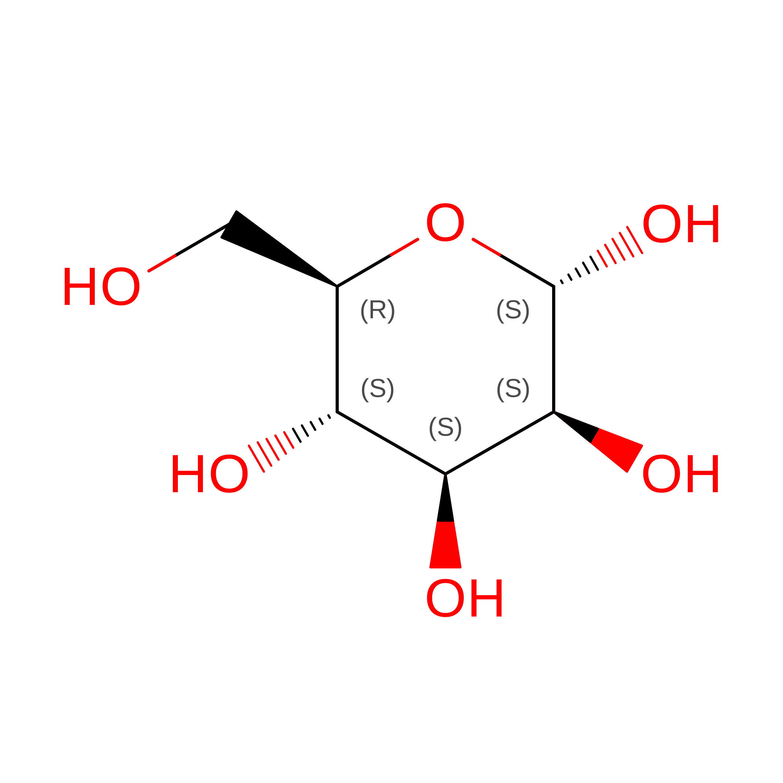
MAN

Query on MAN


Download:Ideal Coordinates CCD File
P [auth C]alpha-D-mannopyranose
C6 H12 O6
WQZGKKKJIJFFOK-PQMKYFCFSA-N
EDO

Query on EDO


Download:Ideal Coordinates CCD File
H [auth A]
I [auth A]
J [auth A]
K [auth B]
L [auth B]
H [auth A],
I [auth A],
J [auth A],
K [auth B],
L [auth B],
M [auth C],
N [auth C],
O [auth C],
Q [auth D],
R [auth D],
S [auth E],
V [auth E]
1,2-ETHANEDIOL
C2 H6 O2
LYCAIKOWRPUZTN-UHFFFAOYSA-N
Modified Residues  1 Unique
IDChains TypeFormula2D DiagramParent
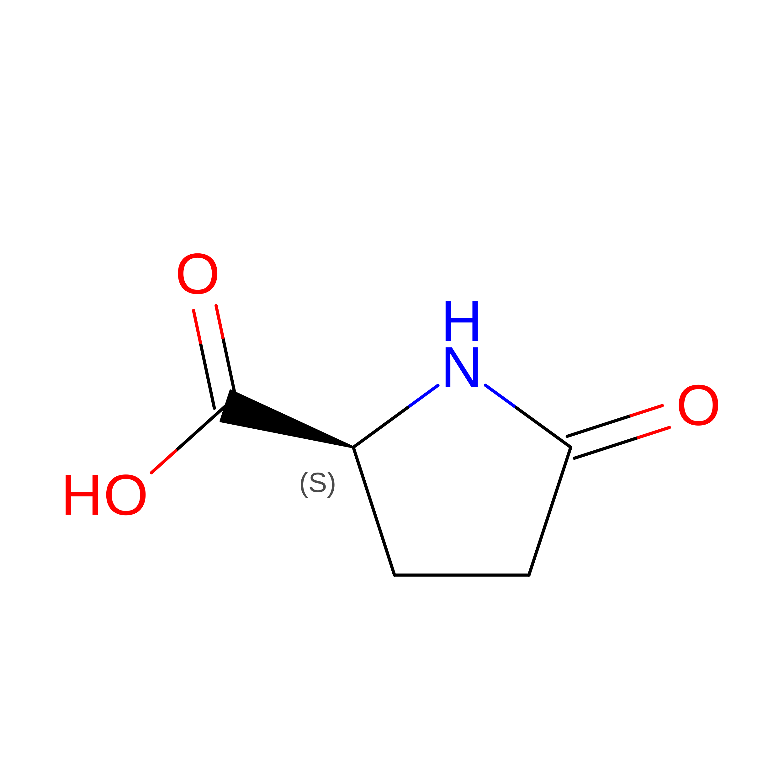
PCA
Query on PCA
E, F
L-PEPTIDE LINKINGC5 H7 N O3GLN

Experimental Data & Validation

Experimental Data

  • Method: X-RAY DIFFRACTION
  • Resolution: 1.56 Å
  • R-Value Free:  0.211 (Depositor), 0.210 (DCC) 
  • R-Value Work:  0.181 (Depositor), 0.180 (DCC) 
  • R-Value Observed: 0.181 (Depositor) 
Space Group: P 1 21 1
Unit Cell:
Length ( Å )Angle ( ˚ )
a = 50.43α = 90
b = 210.6β = 105.561
c = 78.06γ = 90
Software Package:
Software NamePurpose
PHENIXrefinement
XDSdata reduction
XSCALEdata scaling
PHASERphasing

Structure Validation

View Full Validation Report


Ligand Structure Quality Assessment 


Entry History 

& Funding Information

Deposition Data


Funding OrganizationLocationGrant Number
Not funded--

Revision History  (Full details and data files)

  • Version 1.0: 2025-12-10
    Type: Initial release
  • Version 1.1: 2025-12-31
    Changes: Structure summary
  • Version 1.2: 2026-02-18
    Changes: Database references