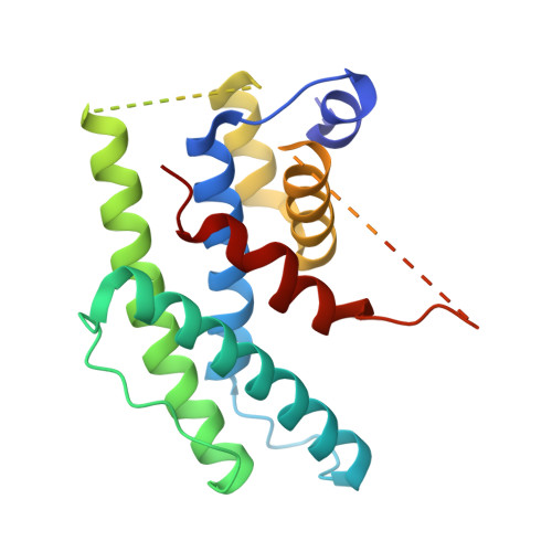
9PHH | pdb_00009phh

TTLL5 CID-RPGR BD Complex


Experimental Data Snapshot

  • Method: X-RAY DIFFRACTION
  • Resolution: 2.80 Å
  • R-Value Free: 
    0.289 (Depositor), 0.289 (DCC) 
  • R-Value Work: 
    0.258 (Depositor), 0.258 (DCC) 
  • R-Value Observed: 
    0.261 (Depositor) 

Starting Model: in silico
View more details

wwPDB Validation 3D Report Full Report

Validation slider image for 9PHH

This is version 1.1 of the entry. See complete history. 

Literature

Insights into retinal disease and non-tubulin glutamylation from a RPGR-TTLL5 complex structure.

Park, J.H., Liu, R.J.Y., Sun, X., Mahalingan, K.K., Hiriyanna, S., Li, T., Roll-Mecak, A.

(2026) J Cell Biol 225

  • DOI: https://doi.org/10.1083/jcb.202508020
  • Primary Citation Related Structures: 
    9PHH

  • PubMed Abstract: 

    Mutations in retinitis pigmentosa GTPase regulator (RPGR) cause photoreceptor degeneration, vision loss, and eventual blindness. RPGR function requires glutamylation by tubulin tyrosine ligase-like 5 (TTLL5) whose mutation is also linked to severe forms of retinal degeneration. How TTLL5 targets RPGR and how mutations in either protein cause disease are unknown. Here we report the 2.8-Å X-ray crystal structure of the coactivator interacting domain (CID) of human TTLL5 in complex with the RPGR C terminus, both required for glutamylation. The RPGR C terminus forms a helix that intercalates through aromatic interactions into the CID helical bundle of novel fold. Interfacial residues are mutated in retinitis pigmentosa, as well as macular degeneration of unknown etiology. Key mutations at this interface abolish RPGR-TTLL5 interaction in vitro and RPGR glutamylation in mouse photoreceptors. Our work reveals mechanisms of non-tubulin substrate recognition by TTLL glutamylases, increasingly recognized as broad regulators of the proteome, and sheds light on mechanisms of disease associated with TTLL5 and RPGR mutations.


  • Organizational Affiliation: 
    • Cell Biology and Biophysics Unit, National Institute of Neurological Disorders and Stroke , Bethesda, MD, USA.

Macromolecule Content 

  • Total Structure Weight: 23.75 kDa 
  • Atom Count: 1,223 
  • Modeled Residue Count: 164 
  • Deposited Residue Count: 209 
  • Unique protein chains: 1

Macromolecules

Find similar proteins by:|  3D Structure
Entity ID: 1
MoleculeChains  Sequence LengthOrganismDetailsImage
Tubulin polyglutamylase TTLL5, Isoform 6 of X-linked retinitis pigmentosa GTPase regulator chimera209Homo sapiensMutation(s): 0 
Gene Names: TTLL5, KIAA0998, STAMP, RPGR, RP3, XLRP3
EC: 6.3.2
UniProt & NIH Common Fund Data Resources
Find proteins for Q6EMB2 (Homo sapiens)
Explore Q6EMB2 
Go to UniProtKB:  Q6EMB2
PHAROS:  Q6EMB2
GTEx:  ENSG00000119685 
Entity Groups
Sequence Clusters30% Identity50% Identity70% Identity90% Identity95% Identity100% Identity
UniProt GroupQ6EMB2
Sequence Annotations
Expand
Reference Sequence

Experimental Data & Validation

Experimental Data

  • Method: X-RAY DIFFRACTION
  • Resolution: 2.80 Å
  • R-Value Free:  0.289 (Depositor), 0.289 (DCC) 
  • R-Value Work:  0.258 (Depositor), 0.258 (DCC) 
  • R-Value Observed: 0.261 (Depositor) 
Space Group: P 41 21 2
Unit Cell:
Length ( Å )Angle ( ˚ )
a = 62.906α = 90
b = 62.906β = 90
c = 166.631γ = 90
Software Package:
Software NamePurpose
PHENIXrefinement
XDSdata reduction
SCALAdata scaling
PHASERphasing

Structure Validation

View Full Validation Report


Entry History 

& Funding Information

Deposition Data


Funding OrganizationLocationGrant Number
National Institutes of Health/National Institute of Neurological Disorders and Stroke (NIH/NINDS)United States1ZIA NS003163-08

Revision History  (Full details and data files)

  • Version 1.0: 2026-04-15
    Type: Initial release
  • Version 1.1: 2026-10-28
    Changes: Database references