9PE4 | pdb_00009pe4

Structure of beta-1,3-glucan synthase from Saccharomyces cerevisiae (ScFks1) at the catalytically less relevant L2 state

  • Classification: MEMBRANE PROTEIN
  • Organism(s): Saccharomyces cerevisiae
  • Mutation(s): No 

  • Deposited: 2025-07-01 Released: 2026-04-22 
  • Deposition Author(s): Ren, Z., Lee, S.Y.
  • Funding Organization(s): National Institutes of Health/National Institute Of Allergy and Infectious Diseases (NIH/NIAID)

Experimental Data Snapshot

  • Method: ELECTRON MICROSCOPY
  • Resolution: 2.69 Å
  • Aggregation State: PARTICLE 
  • Reconstruction Method: SINGLE PARTICLE 

wwPDB Validation 3D Report Full Report

Validation slider image for 9PE4

This is version 1.1 of the entry. See complete history

Literature

Structural basis of fungal beta-1,3-glucan synthase inhibition by caspofungin.

Ren, Z.Chhetri, A.Liu, C.Offner, S.Sharma, K.Borgnia, M.J.Im, W.Yokoyama, K.Lee, S.Y.

(2026) Nature 

  • DOI: https://doi.org/10.1038/s41586-026-10409-7
  • Primary Citation Related Structures: 
    9PE1, 9PE2, 9PE3, 9PE4, 9PE5, 9ZTC

  • PubMed Abstract: 

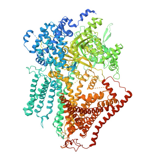
    Invasive fungal infections pose life-threatening risks to the increasing population of immunocompromised patients 1,2 . Treatment remains challenging due to limited antifungal drugs and increasing resistance. β-1,3-D-glucan synthase (GS), comprising the catalytic Fks1 and the regulatory small GTPase, Rho1 (refs. 3,4 ), is the target of clinically important echinocandin antifungals. Despite recent studies 5-7 , the mechanisms of GS catalysis, Rho1 regulation and echinocandin inhibition and resistance remain elusive. Here we present cryo-electron microscopy structures of native Saccharomyces cerevisiae Fks1 (ScFks1) solved under catalytically relevant conditions, revealing its interactions with the antifungal caspofungin (CFN), glucan product from the translocation channel and Rho1. CFN forms a ternary complex with nascent glucan and Fks1 at the membrane-protein interface, suggesting an unexpected role of CFN in stalling polymer translocation. Our echinocandin-resistant S643P structure suggests a resistance mechanism: the substitution destabilizes CFN and glucan binding through both allosteric structural perturbation and direct steric clash. Rho1 binding induces active site rearrangements essential for catalysis, including that of the 'latch loop' for donor substrate coordination. Furthermore, we identify YMR295C as an auxiliary subunit. These findings elucidate the mechanisms of GS-mediated glucan synthesis and its inhibition and resistance by echinocandins, laying the groundwork for rational antifungal design.


  • Organizational Affiliation
    • Department of Biochemistry, Duke University School of Medicine, Durham, NC, USA.

Macromolecule Content 

  • Total Structure Weight: 250.21 kDa 
  • Atom Count: 13,050 
  • Modeled Residue Count: 1,577 
  • Deposited Residue Count: 2,128 
  • Unique protein chains: 2

Macromolecules

Find similar proteins by:|  3D Structure
Entity ID: 1
MoleculeChains  Sequence LengthOrganismDetailsImage
1,3-beta-glucan synthase component FKS11,931Saccharomyces cerevisiaeMutation(s): 0 
EC: 2.4.1.34
UniProt
Find proteins for P38631 (Saccharomyces cerevisiae (strain ATCC 204508 / S288c))
Explore P38631 
Go to UniProtKB:  P38631
Entity Groups
Sequence Clusters30% Identity50% Identity70% Identity90% Identity95% Identity100% Identity
UniProt GroupP38631
Sequence Annotations
Expand
Reference Sequence
Find similar proteins by:|  3D Structure
Entity ID: 2
MoleculeChains  Sequence LengthOrganismDetailsImage
YMR295CB [auth G]197Saccharomyces cerevisiaeMutation(s): 0 
UniProt
Find proteins for Q03559 (Saccharomyces cerevisiae (strain ATCC 204508 / S288c))
Explore Q03559 
Go to UniProtKB:  Q03559
Entity Groups
Sequence Clusters30% Identity50% Identity70% Identity90% Identity95% Identity100% Identity
UniProt GroupQ03559
Sequence Annotations
Expand
Reference Sequence

Oligosaccharides

Help  
Entity ID: 3
MoleculeChains Length2D Diagram GlycosylationD Interactions
beta-D-mannopyranose-(1-4)-2-acetamido-2-deoxy-beta-D-glucopyranose-(1-4)-2-acetamido-2-deoxy-beta-D-glucopyranoseC [auth D]3N/A
Glycosylation Resources
GlyTouCan: G15407YE
GlyCosmos: G15407YE
GlyGen: G15407YE

Small Molecules

Ligands 3 Unique
IDChains Name / Formula / InChI Key2D Diagram3D Interactions
LMN

Query on LMN


Download:Ideal Coordinates CCD File
L [auth A],
M [auth A]
Lauryl Maltose Neopentyl Glycol
C47 H88 O22
MADJBYLAYPCCOO-XYPZXBMFSA-N
3PE

Query on 3PE


Download:Ideal Coordinates CCD File
H [auth A]1,2-Distearoyl-sn-glycerophosphoethanolamine
C41 H82 N O8 P
LVNGJLRDBYCPGB-LDLOPFEMSA-N
Y01

Query on Y01


Download:Ideal Coordinates CCD File
D [auth A]
E [auth A]
F [auth A]
G [auth A]
I [auth A]
D [auth A],
E [auth A],
F [auth A],
G [auth A],
I [auth A],
J [auth A],
K [auth A]
CHOLESTEROL HEMISUCCINATE
C31 H50 O4
WLNARFZDISHUGS-MIXBDBMTSA-N

Experimental Data & Validation

Experimental Data

  • Method: ELECTRON MICROSCOPY
  • Resolution: 2.69 Å
  • Aggregation State: PARTICLE 
  • Reconstruction Method: SINGLE PARTICLE 
EM Software:
TaskSoftware PackageVersion
MODEL REFINEMENTPHENIX1.20.1_4487:
RECONSTRUCTIONcryoSPARC

Structure Validation

View Full Validation Report


Entry History 

& Funding Information

Deposition Data


Funding OrganizationLocationGrant Number
National Institutes of Health/National Institute Of Allergy and Infectious Diseases (NIH/NIAID)United StatesR01AI170906

Revision History  (Full details and data files)

  • Version 1.0: 2026-04-22
    Type: Initial release
  • Version 1.1: 2026-05-06
    Changes: Data collection, Database references