9P9E | pdb_00009p9e

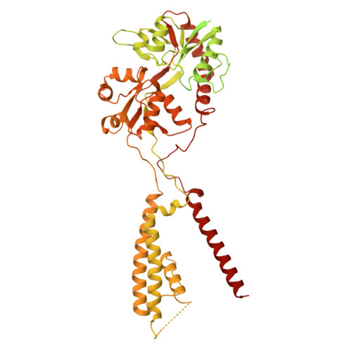
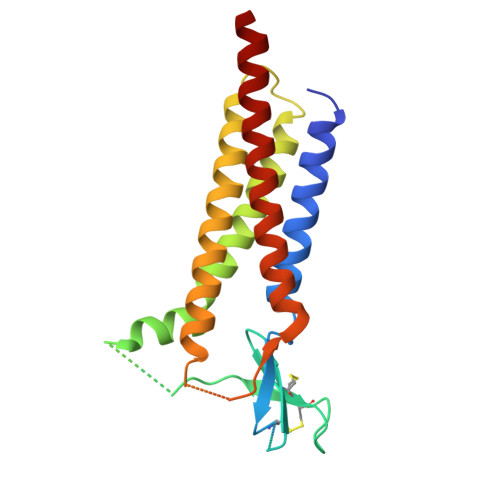
Active substate 3 of the GluA4 homotetramer.

  • Classification: MEMBRANE PROTEIN
  • Organism(s): Rattus norvegicus, Mus musculus
  • Expression System: Mus musculus, Homo sapiens
  • Mutation(s): No 

  • Deposited: 2025-06-24 Released: 2026-01-28 
  • Deposition Author(s): Hale, W.D., Huganir, R.L., Twomey, E.C.
  • Funding Organization(s): National Institutes of Health/National Institute of Mental Health (NIH/NIMH), National Institutes of Health/National Institute of Neurological Disorders and Stroke (NIH/NINDS), National Institutes of Health/National Institute of General Medical Sciences (NIH/NIGMS)

Experimental Data Snapshot

  • Method: ELECTRON MICROSCOPY
  • Resolution: 3.82 Å
  • Aggregation State: PARTICLE 
  • Reconstruction Method: SINGLE PARTICLE 

wwPDB Validation 3D Report Full Report

Validation slider image for 9P9E

This is version 1.1 of the entry. See complete history. 

Literature

Structural basis for activation and conformational plasticity of the GluA4 AMPA receptor.

Hale, W.D., Wang, H., Huganir, R.L., Twomey, E.C.

(2026) Nat Commun 17

  • DOI: https://doi.org/10.1038/s41467-026-68953-9
  • Primary Citation Related Structures: 
    9P9B, 9P9C, 9P9D, 9P9E, 9P9F, 9P9G

  • PubMed Abstract: 

    AMPA-subtype glutamate receptors (AMPARs) mediate excitatory synaptic transmission. AMPAR ion channels exhibit multiple subconductance states that tune neuronal responses to glutamate. GluA4 is the rarest subunit in the brain but is enriched in interneurons and the cerebellum. Rising evidence points to GluA4 AMPARs in the development of neurological diseases, but the structural mechanisms of GluA4 function remain enigmatic. Here, we show the distinct features of GluA4 that tune AMPAR function. We find that GluA4 AMPARs have a canonical "Y" shaped architecture where local dimer pairs are domain-swapped between the amino terminal domain (NTD) and ligand binding domain (LBD), both of which comprise the extracellular domain. All four LBDs are glutamate bound yet open the GluA4 ion channel by asymmetric hinging in all four channel helices. We observe that the glutamate-saturated LBD has conformational plasticity, which tunes the ion channel gate below. These data provide a framework for understanding channel subconductance, outline the distinct properties of GluA4, expand our understanding of conformational plasticity in AMPARs, and will inform therapeutic design.


  • Organizational Affiliation: 
    • Solomon H. Snyder Department of Neuroscience, Johns Hopkins University School of Medicine, Baltimore, MD, USA.

Macromolecule Content 

  • Total Structure Weight: 474.97 kDa 
  • Atom Count: 18,100 
  • Modeled Residue Count: 2,304 
  • Deposited Residue Count: 4,220 
  • Unique protein chains: 2

Macromolecules

Find similar proteins by:|  3D Structure
Entity ID: 1
MoleculeChains  Sequence LengthOrganismDetailsImage
Isoform 2 of Glutamate receptor 4
A, B, C, D
846Rattus norvegicusMutation(s): 0 
Gene Names: Gria4, Glur4
UniProt
Find proteins for P19493 (Rattus norvegicus)
Explore P19493 
Go to UniProtKB:  P19493
Entity Groups
Sequence Clusters30% Identity50% Identity70% Identity90% Identity95% Identity100% Identity
UniProt GroupP19493
Sequence Annotations
Expand
Reference Sequence
Find similar proteins by:|  3D Structure
Entity ID: 2
MoleculeChains  Sequence LengthOrganismDetailsImage
Voltage-dependent calcium channel gamma-2 subunitE [auth H],
F,
G,
H [auth E]
209Mus musculusMutation(s): 0 
Gene Names: Cacng2, Stg
UniProt & NIH Common Fund Data Resources
Find proteins for O88602 (Mus musculus)
Explore O88602 
Go to UniProtKB:  O88602
IMPC:  MGI:1316660
Entity Groups
Sequence Clusters30% Identity50% Identity70% Identity90% Identity95% Identity100% Identity
UniProt GroupO88602
Sequence Annotations
Expand
Reference Sequence

Small Molecules

Ligands 2 Unique
IDChains Name / Formula / InChI Key2D Diagram3D Interactions
CYZ

Query on CYZ


Download:Ideal Coordinates CCD File
J [auth A],
L [auth B],
M [auth B],
P [auth D]
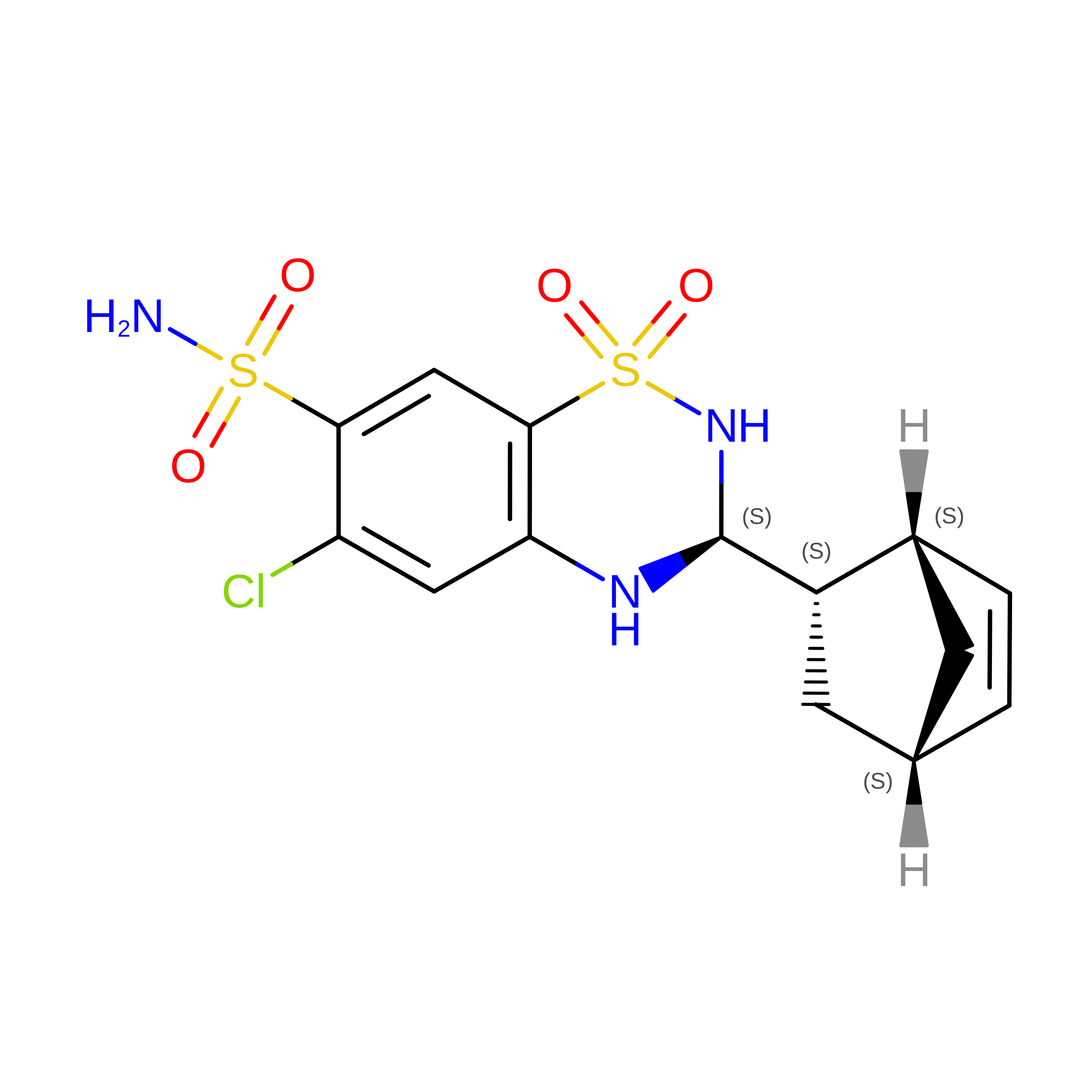
CYCLOTHIAZIDE
C14 H16 Cl N3 O4 S2
BOCUKUHCLICSIY-KSCJFIISSA-N
GLU

Query on GLU


Download:Ideal Coordinates CCD File
I [auth A],
K [auth B],
N [auth C],
O [auth D]
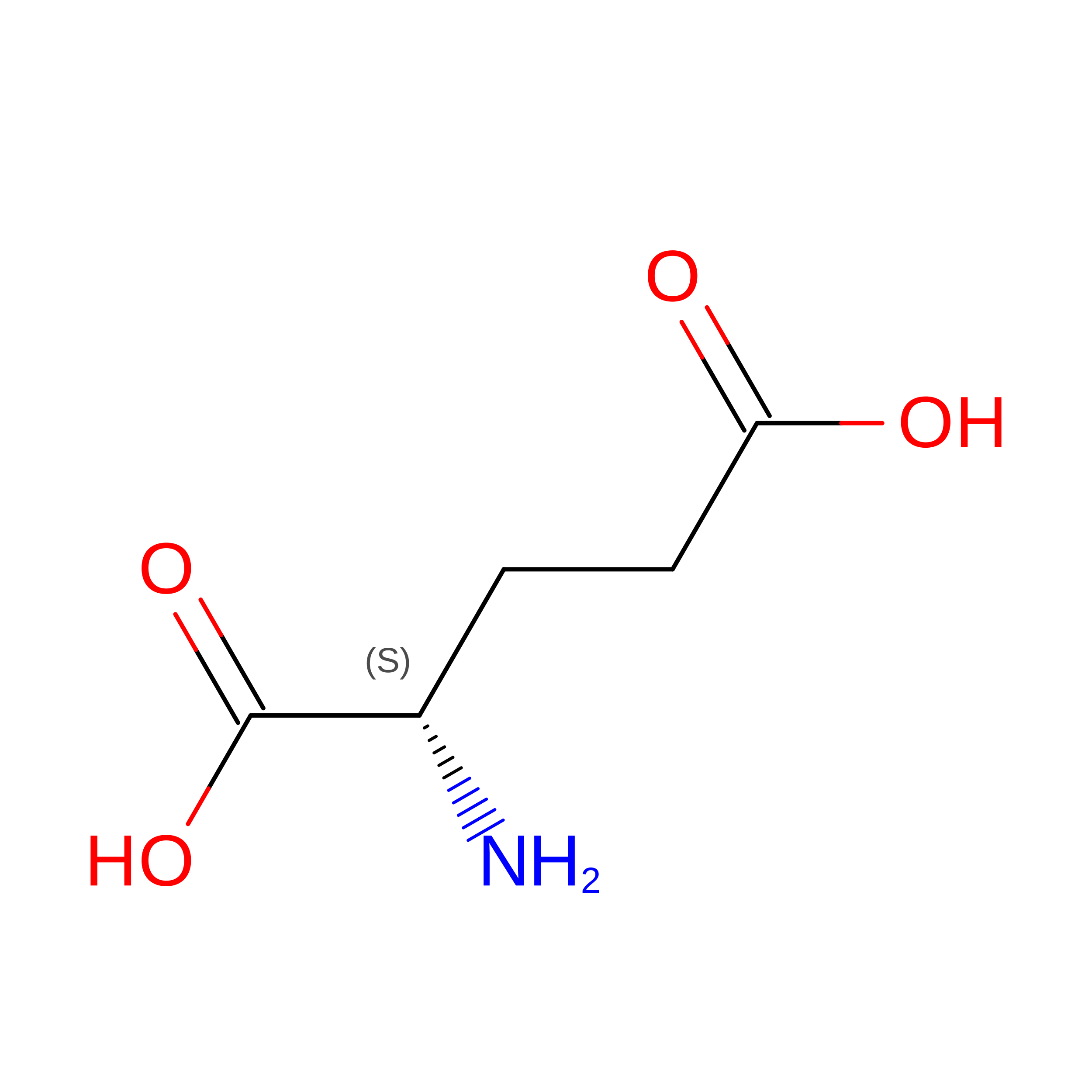
GLUTAMIC ACID
C5 H9 N O4
WHUUTDBJXJRKMK-VKHMYHEASA-N
Binding Affinity Annotations 
IDSourceBinding Affinity
CYZ BindingDB:  9P9E EC50: 3800 (nM) from 1 assay(s)

Experimental Data & Validation

Experimental Data

  • Method: ELECTRON MICROSCOPY
  • Resolution: 3.82 Å
  • Aggregation State: PARTICLE 
  • Reconstruction Method: SINGLE PARTICLE 
EM Software:
TaskSoftware PackageVersion
MODEL REFINEMENTPHENIX1.21.2_5419
RECONSTRUCTIONcryoSPARC

Structure Validation

View Full Validation Report


Entry History 

& Funding Information

Deposition Data


Funding OrganizationLocationGrant Number
National Institutes of Health/National Institute of Mental Health (NIH/NIMH)United StatesK99MH132811
National Institutes of Health/National Institute of Neurological Disorders and Stroke (NIH/NINDS)United StatesR37NS036715
National Institutes of Health/National Institute of General Medical Sciences (NIH/NIGMS)United StatesR35GM154904

Revision History  (Full details and data files)

  • Version 1.0: 2026-01-28
    Type: Initial release
  • Version 1.1: 2026-09-23
    Changes: Data collection, Database references