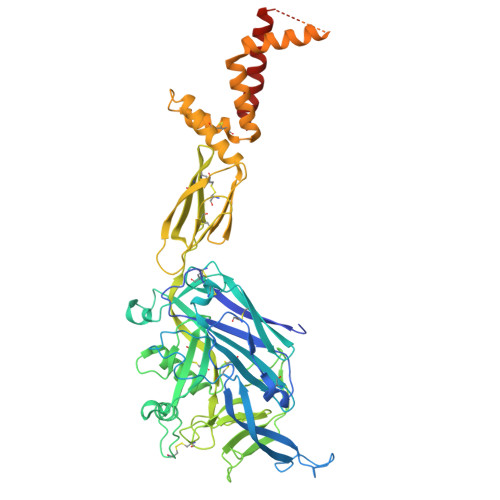
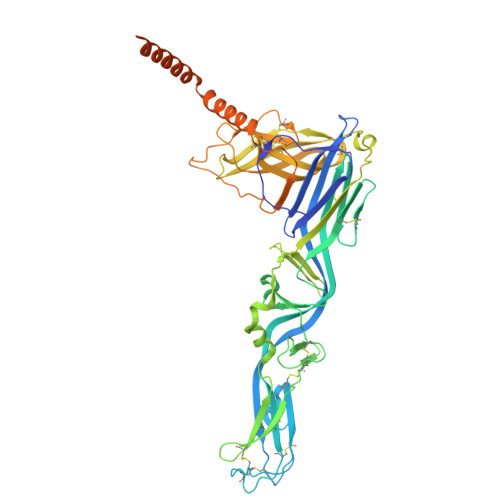
9P3I | pdb_00009p3i

High-resolution in situ ANDV single tetramer structure


Experimental Data Snapshot

  • Method: ELECTRON MICROSCOPY
  • Resolution: 2.35 Å
  • Aggregation State: PARTICLE 
  • Reconstruction Method: SINGLE PARTICLE 

wwPDB Validation 3D Report Full Report

Validation slider image for 9P3I

This is version 1.3 of the entry. See complete history. 

Literature

High-resolution in situ structures of hantavirus glycoprotein tetramers.

Guo, L., McFadden, E., Slough, M.M., Stone, E.T., Berrigan, J., Mittler, E., Hatzakis, K., Hinkley, T., Kain, H.S., Ke, Z., Warner, N.L., Erasmus, J.H., Chandran, K., McLellan, J.S.

(2026) Cell 189: 2731

  • DOI: https://doi.org/10.1016/j.cell.2026.01.030
  • Primary Citation Related Structures: 
    9P3I, 9P3L, 9P3M, 9P3X, 9P3Y

  • PubMed Abstract: 

    New World hantaviruses cause severe infections in humans. Previous structural studies have advanced our understanding of hantavirus glycoprotein architecture; however, the lack of high-resolution structures of the glycoprotein tetramer and its lattice organization has limited mechanistic insights into viral assembly and entry. Here, we leveraged a virus-like particle (VLP) system to establish a cryo-electron microscopy workflow for lattice-forming viral glycoproteins. This enabled the determination of a 2.35 Å resolution structure of the membrane-embedded Andes virus (ANDV) glycoprotein tetramer as well as the structures of dimers of tetramers and a complex with antibody ADI-65534. These structures reveal previously uncharacterized features of glycoprotein organization, stability, and pH sensing. The immunization of mice with self-amplifying replicon RNA (repRNA) encoding ANDV-VLPs elicited high levels of glycoprotein-binding antibodies but equivalent titers of neutralizing antibodies compared with the repRNA-encoded native ANDV glycoprotein complex. These findings advance our understanding of hantavirus glycoprotein assemblies, laying a foundation for structure-based vaccine design.


  • Organizational Affiliation: 
    • Department of Molecular Biosciences, The University of Texas at Austin, Austin, TX 78712, USA.

Macromolecule Content 

  • Total Structure Weight: 535.48 kDa 
  • Atom Count: 31,608 
  • Modeled Residue Count: 3,964 
  • Deposited Residue Count: 4,752 
  • Unique protein chains: 2

Macromolecules

Find similar proteins by:|  3D Structure
Entity ID: 1
MoleculeChains  Sequence LengthOrganismDetailsImage
Glycoprotein N
A, C, E, G
651Orthohantavirus andesenseMutation(s): 1 
Gene Names: GP, ADT63_77597gpM, ADT63_77598gpM
UniProt
Find proteins for Q9E006 (Andes orthohantavirus)
Explore Q9E006 
Go to UniProtKB:  Q9E006
Entity Groups
Sequence Clusters30% Identity50% Identity70% Identity90% Identity95% Identity100% Identity
UniProt GroupQ9E006
Glycosylation
Glycosylation Sites: 3
Sequence Annotations
Expand
Reference Sequence
Find similar proteins by:|  3D Structure
Entity ID: 2
MoleculeChains  Sequence LengthOrganismDetailsImage
Glycoprotein C
B, D, F, H
537Orthohantavirus andesenseMutation(s): 1 
Gene Names: GP, ADT63_77597gpM, ADT63_77598gpM
UniProt
Find proteins for Q9E006 (Andes orthohantavirus)
Explore Q9E006 
Go to UniProtKB:  Q9E006
Entity Groups
Sequence Clusters30% Identity50% Identity70% Identity90% Identity95% Identity100% Identity
UniProt GroupQ9E006
Glycosylation
Glycosylation Sites: 1
Sequence Annotations
Expand
Reference Sequence

Oligosaccharides

Help  
Entity ID: 3
MoleculeChains Length2D Diagram GlycosylationD Interactions
alpha-D-mannopyranose-(1-3)-[alpha-D-mannopyranose-(1-6)]beta-D-mannopyranose-(1-4)-2-acetamido-2-deoxy-beta-D-glucopyranose-(1-4)-2-acetamido-2-deoxy-beta-D-glucopyranose
I, M, Q, U
5N-Glycosylation
Glycosylation Resources
GlyTouCan: G22768VO
GlyCosmos: G22768VO
GlyGen: G22768VO
Entity ID: 4
MoleculeChains Length2D Diagram GlycosylationD Interactions
alpha-D-mannopyranose-(1-3)-beta-D-mannopyranose-(1-4)-2-acetamido-2-deoxy-beta-D-glucopyranose-(1-4)-2-acetamido-2-deoxy-beta-D-glucopyranose
J, N, R, V
4N-Glycosylation
Glycosylation Resources
GlyTouCan: G81315DD
GlyCosmos: G81315DD
GlyGen: G81315DD
Entity ID: 5
MoleculeChains Length2D Diagram GlycosylationD Interactions
alpha-D-mannopyranose-(1-6)-beta-D-mannopyranose-(1-4)-2-acetamido-2-deoxy-beta-D-glucopyranose-(1-4)-2-acetamido-2-deoxy-beta-D-glucopyranose
K, O, S, W
4N-Glycosylation
Glycosylation Resources
GlyTouCan: G22573RC
GlyCosmos: G22573RC
GlyGen: G22573RC
Entity ID: 6
MoleculeChains Length2D Diagram GlycosylationD Interactions
2-acetamido-2-deoxy-beta-D-glucopyranose-(1-4)-2-acetamido-2-deoxy-beta-D-glucopyranose
L, P, T, X
2N-Glycosylation
Glycosylation Resources
GlyTouCan: G42666HT
GlyCosmos: G42666HT
GlyGen: G42666HT

Experimental Data & Validation

Experimental Data

  • Method: ELECTRON MICROSCOPY
  • Resolution: 2.35 Å
  • Aggregation State: PARTICLE 
  • Reconstruction Method: SINGLE PARTICLE 
EM Software:
TaskSoftware PackageVersion
MODEL REFINEMENTPHENIX1.21.2_5419
RECONSTRUCTIONcryoSPARC4.6.2

Structure Validation

View Full Validation Report


Entry History 

& Funding Information

Deposition Data


Funding OrganizationLocationGrant Number
National Institutes of Health/National Institute Of Allergy and Infectious Diseases (NIH/NIAID)United StatesU19 AI181977

Revision History  (Full details and data files)

  • Version 1.0: 2025-07-30
    Type: Initial release
  • Version 1.1: 2025-11-19
    Changes: Author supporting evidence, Data collection
  • Version 1.2: 2026-03-25
    Changes: Data collection, Database references
  • Version 1.3: 2026-05-13
    Changes: Data collection, Database references