9P1I | pdb_00009p1i

Atomic structure of vibrio effector fragment VopV bound to Beta-cytoplasmic/gamma1-cytoplasmic F-actin


Experimental Data Snapshot

  • Method: ELECTRON MICROSCOPY
  • Resolution: 3.00 Å
  • Aggregation State: FILAMENT 
  • Reconstruction Method: HELICAL 

wwPDB Validation 3D Report Full Report

Validation slider image for 9P1I

This is version 1.1 of the entry. See complete history

Literature

Actin isoform-specific interactions revealed by Vibrio VopV actin-binding repeats.

Kudryashova, E.Kreutzberger, M.A.B.Niedzialkowska, E.Dong, S.Kudryashov, D.S.Egelman, E.H.

(2025) Proc Natl Acad Sci U S A 122: e2523856122-e2523856122

  • DOI: https://doi.org/10.1073/pnas.2523856122
  • Primary Citation Related Structures: 
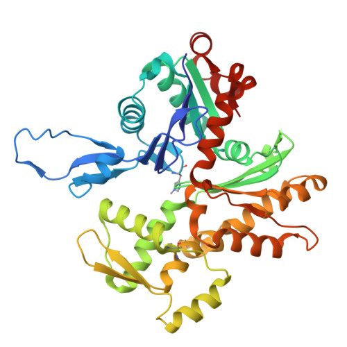
    9P1I, 9P3D

  • PubMed Abstract: 

    Despite an evolutionary separation of over 300 Mya, there are no amino acid substitutions in certain actin isoforms from reptiles to mammals. What divergence that does exist between different actin isoforms is primarily tissue-specific, rather than species-specific. Sorting of actin isoforms into distinct cellular compartments is believed to be controlled by actin-binding proteins (ABPs), but little is known about how ABPs can differentiate between actin isoforms. We show that the actin-binding repeat (ABR) of the Vibrio parahaemolyticus effector VopV binds to cytoplasmic actin in a unique mode with a low nanomolar affinity, over a thousand times stronger than to muscle actin. Actin mutagenesis and cryo-EM reconstructions reveal that isoform-specific residues of previously unassigned function deep in the cleft between the two actin protofilament strands determine this selectivity. These results suggest a mechanism of highly selective, isoform-specific interactions between actin and its partners, and have broad implications for understanding the evolution of actin. Furthermore, our findings have implications in the pathogenesis of V. parahaemolyticus, whose invasion of intestinal epithelial cells relies on the interaction of VopV with cytoplasmic F-actin.


  • Organizational Affiliation
    • Department of Chemistry and Biochemistry, The Ohio State University, Columbus, OH 43210.

Macromolecule Content 

  • Total Structure Weight: 3,440.18 kDa 
  • Atom Count: 39,624 
  • Modeled Residue Count: 5,028 
  • Deposited Residue Count: 33,324 
  • Unique protein chains: 2

Macromolecules

Find similar proteins by:|  3D Structure
Entity ID: 1
MoleculeChains  Sequence LengthOrganismDetailsImage
Actin, cytoplasmic 1375Homo sapiensMutation(s): 0 
Gene Names: ACTB
EC: 3.6.4
UniProt & NIH Common Fund Data Resources
Find proteins for P60709 (Homo sapiens)
Explore P60709 
Go to UniProtKB:  P60709
PHAROS:  P60709
GTEx:  ENSG00000075624 
Entity Groups
Sequence Clusters30% Identity50% Identity70% Identity90% Identity95% Identity100% Identity
UniProt GroupP60709
Sequence Annotations
Expand
Reference Sequence
Find similar proteins by:|  3D Structure
Entity ID: 2
MoleculeChains  Sequence LengthOrganismDetailsImage
VopV2,402Vibrio parahaemolyticusMutation(s): 0 
Gene Names: MAVP-QPI_00061
UniProt
Find proteins for A0A250E4R0 (Vibrio parahaemolyticus)
Explore A0A250E4R0 
Go to UniProtKB:  A0A250E4R0
Entity Groups
Sequence Clusters30% Identity50% Identity70% Identity90% Identity95% Identity100% Identity
UniProt GroupA0A250E4R0
Sequence Annotations
Expand
Reference Sequence

Small Molecules

Ligands 2 Unique
IDChains Name / Formula / InChI Key2D Diagram3D Interactions
ANP

Query on ANP


Download:Ideal Coordinates CCD File
BA [auth R]
DA [auth S]
FA [auth T]
HA [auth U]
JA [auth V]
BA [auth R],
DA [auth S],
FA [auth T],
HA [auth U],
JA [auth V],
LA [auth W],
NA [auth X],
PA [auth Y],
RA [auth Z],
TA [auth a],
VA [auth b],
Z [auth Q]
PHOSPHOAMINOPHOSPHONIC ACID-ADENYLATE ESTER
C10 H17 N6 O12 P3
PVKSNHVPLWYQGJ-KQYNXXCUSA-N
MG

Query on MG


Download:Ideal Coordinates CCD File
AA [auth R]
CA [auth S]
EA [auth T]
GA [auth U]
IA [auth V]
AA [auth R],
CA [auth S],
EA [auth T],
GA [auth U],
IA [auth V],
KA [auth W],
MA [auth X],
OA [auth Y],
QA [auth Z],
SA [auth a],
UA [auth b],
Y [auth Q]
MAGNESIUM ION
Mg
JLVVSXFLKOJNIY-UHFFFAOYSA-N
Modified Residues  1 Unique
IDChains TypeFormula2D DiagramParent
HIC
Query on HIC
A [auth Q]
B [auth R]
C [auth S]
D [auth T]
E [auth U]
A [auth Q],
B [auth R],
C [auth S],
D [auth T],
E [auth U],
F [auth V],
G [auth W],
H [auth X],
I [auth Y],
J [auth Z],
K [auth a],
L [auth b]
L-PEPTIDE LINKINGC7 H11 N3 O2HIS

Experimental Data & Validation

Experimental Data

  • Method: ELECTRON MICROSCOPY
  • Resolution: 3.00 Å
  • Aggregation State: FILAMENT 
  • Reconstruction Method: HELICAL 
EM Software:
TaskSoftware PackageVersion
RECONSTRUCTIONcryoSPARC4.51

Structure Validation

View Full Validation Report


Entry History 

& Funding Information

Deposition Data


Funding OrganizationLocationGrant Number
National Institutes of Health/National Institute of General Medical Sciences (NIH/NIGMS)United StatesR35 GM122510
National Institutes of Health/National Institute of General Medical Sciences (NIH/NIGMS)United StatesR01 GM114666

Revision History  (Full details and data files)

  • Version 1.0: 2025-11-19
    Type: Initial release
  • Version 1.1: 2025-12-10
    Changes: Data collection, Database references