9OUM | pdb_00009oum

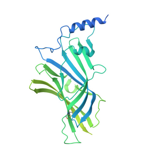
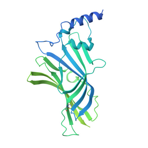
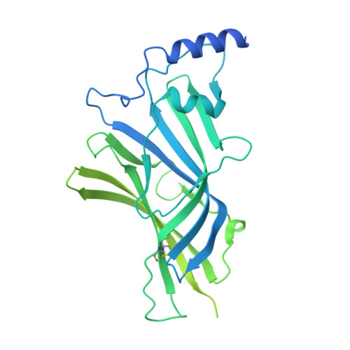
Native GABA-A receptor from rat cerebella, beta2-alpha1-beta1-alpha6-gamma2 subtype, in complex with GABA and PZ-II-029


Experimental Data Snapshot

  • Method: ELECTRON MICROSCOPY
  • Resolution: 2.90 Å
  • Aggregation State: PARTICLE 
  • Reconstruction Method: SINGLE PARTICLE 

wwPDB Validation 3D Report Full Report

Validation slider image for 9OUM

This is version 1.0 of the entry. See complete history

Literature

Molecular assemblies and pharmacology of cerebellar GABA A receptors.

Sun, C.Jahncke, J.N.Wright, K.M.Gouaux, E.

(2026) Proc Natl Acad Sci U S A 123: e2524504123-e2524504123

  • DOI: https://doi.org/10.1073/pnas.2524504123
  • Primary Citation Related Structures: 
    9OUM, 9OUN, 9OUO, 9OUP, 9OUQ, 9OUR, 9OV4

  • PubMed Abstract: 

    GABA A receptors (GABA A Rs) mediate fast inhibitory neurotransmission in the brain and are assembled from 19 subunit isoforms into multiple pentameric assemblies. Although α1-containing GABA A Rs are broadly expressed and are pharmacologically important, the molecular diversity of native α1-based assemblies in specific brain regions remains incompletely understood. Here, we use immunofluorescence, mass spectrometry, and cryogenic electron microscopy (cryo-EM) to characterize the spatial distribution, subunit composition, and structural architecture of native α1-containing GABA A Rs in the rat cerebellum. Confocal microscopy reveals robust colocalization of α1 and γ2 subunits across cerebellar layers, including prominent labeling at glomerular synapses. Biochemical purification and proteomic analysis identify a range of α, β, and γ subunits, along with abundant α6 and δ subunits. Using cryo-EM and automated subunit identification, we resolve eight α1-containing receptor assemblies, including the first structure of α6-containing receptors. We further determine the binding mode of the α6-selective pyrazoloquinolinone modulator PZ-II-029 at the α + - interface, showing ligand-induced expansion of the entire extracellular domain (ECD). Together, our study defines the structure and subunit composition of the α1-containing cerebellar GABA A Rs and elaborates the molecular interactions between native receptors and pyrazoloquinolinone, thereby laying the groundwork for brain region and subunit-specific pharmacology.


  • Organizational Affiliation
    • Vollum Institute, Oregon Health and Science University, Portland, OR 97239.

Macromolecule Content 

  • Total Structure Weight: 319.24 kDa 
  • Atom Count: 10,660 
  • Modeled Residue Count: 1,277 
  • Deposited Residue Count: 2,758 
  • Unique protein chains: 7

Macromolecules

Find similar proteins by:|  3D Structure
Entity ID: 1
MoleculeChains  Sequence LengthOrganismDetailsImage
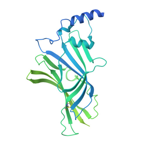
Gamma-aminobutyric acid receptor subunit beta-1A [auth B]474Rattus norvegicusMutation(s): 0 
UniProt
Find proteins for P15431 (Rattus norvegicus)
Explore P15431 
Go to UniProtKB:  P15431
Entity Groups
Sequence Clusters30% Identity50% Identity70% Identity90% Identity95% Identity100% Identity
UniProt GroupP15431
Glycosylation
Glycosylation Sites: 2Go to GlyGen: P15431-1
Sequence Annotations
Expand
Reference Sequence
Find similar proteins by:|  3D Structure
Entity ID: 2
MoleculeChains  Sequence LengthOrganismDetailsImage
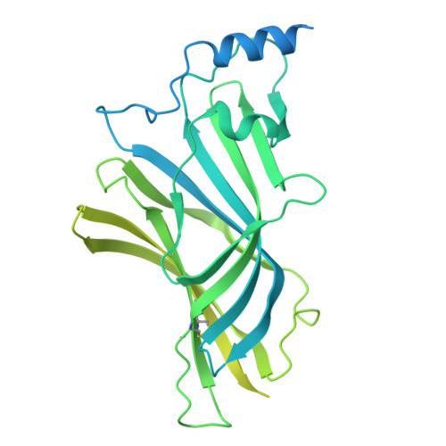
Gamma-aminobutyric acid receptor subunit gamma-2B [auth D]466Rattus norvegicusMutation(s): 0 
UniProt
Find proteins for P18508 (Rattus norvegicus)
Explore P18508 
Go to UniProtKB:  P18508
Entity Groups
Sequence Clusters30% Identity50% Identity70% Identity90% Identity95% Identity100% Identity
UniProt GroupP18508
Glycosylation
Glycosylation Sites: 2Go to GlyGen: P18508-1
Sequence Annotations
Expand
Reference Sequence
Find similar proteins by:|  3D Structure
Entity ID: 3
MoleculeChains  Sequence LengthOrganismDetailsImage
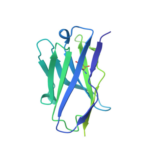
8E3 Fab heavy chainC [auth H]223Mus musculusMutation(s): 0 
Find similar proteins by:|  3D Structure
Entity ID: 4
MoleculeChains  Sequence LengthOrganismDetailsImage
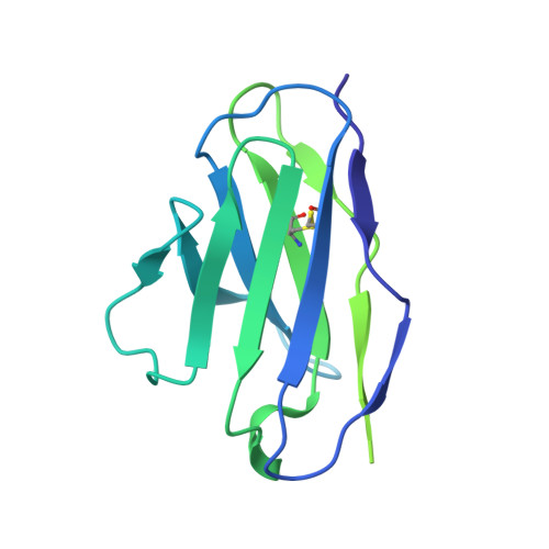
8E3 Fab light chainD [auth L]213Mus musculusMutation(s): 0 
Find similar proteins by:|  3D Structure
Entity ID: 5
MoleculeChains  Sequence LengthOrganismDetailsImage
Gamma-aminobutyric acid receptor subunit alpha-1E [auth A]455Rattus norvegicusMutation(s): 0 
UniProt
Find proteins for P62813 (Rattus norvegicus)
Explore P62813 
Go to UniProtKB:  P62813
Entity Groups
Sequence Clusters30% Identity50% Identity70% Identity90% Identity95% Identity100% Identity
UniProt GroupP62813
Glycosylation
Glycosylation Sites: 1Go to GlyGen: P62813-1
Sequence Annotations
Expand
Reference Sequence
Find similar proteins by:|  3D Structure
Entity ID: 6
MoleculeChains  Sequence LengthOrganismDetailsImage
Gamma-aminobutyric acid receptor subunit beta-2F [auth E]474Rattus norvegicusMutation(s): 0 
UniProt
Find proteins for P63138 (Rattus norvegicus)
Explore P63138 
Go to UniProtKB:  P63138
Entity Groups
Sequence Clusters30% Identity50% Identity70% Identity90% Identity95% Identity100% Identity
UniProt GroupP63138
Glycosylation
Glycosylation Sites: 2Go to GlyGen: P63138-1
Sequence Annotations
Expand
Reference Sequence
Find similar proteins by:|  3D Structure
Entity ID: 7
MoleculeChains  Sequence LengthOrganismDetailsImage
Gamma-aminobutyric acid receptor subunit alpha-6G [auth C]453Rattus norvegicusMutation(s): 0 
UniProt
Find proteins for P30191 (Rattus norvegicus)
Explore P30191 
Go to UniProtKB:  P30191
Entity Groups
Sequence Clusters30% Identity50% Identity70% Identity90% Identity95% Identity100% Identity
UniProt GroupP30191
Glycosylation
Glycosylation Sites: 2Go to GlyGen: P30191-1
Sequence Annotations
Expand
Reference Sequence

Oligosaccharides

Help  
Entity ID: 8
MoleculeChains Length2D Diagram GlycosylationD Interactions
2-acetamido-2-deoxy-beta-D-glucopyranose-(1-4)-2-acetamido-2-deoxy-beta-D-glucopyranoseH [auth F],
J [auth I],
L [auth K],
N
2N-Glycosylation
Glycosylation Resources
GlyTouCan: G42666HT
GlyCosmos: G42666HT
GlyGen: G42666HT
Entity ID: 9
MoleculeChains Length2D Diagram GlycosylationD Interactions
beta-D-mannopyranose-(1-4)-2-acetamido-2-deoxy-beta-D-glucopyranose-(1-4)-2-acetamido-2-deoxy-beta-D-glucopyranoseI [auth G],
M,
O
3N-Glycosylation
Glycosylation Resources
GlyTouCan: G15407YE
GlyCosmos: G15407YE
GlyGen: G15407YE
Entity ID: 10
MoleculeChains Length2D Diagram GlycosylationD Interactions
alpha-D-mannopyranose-(1-2)-alpha-D-mannopyranose-(1-3)-[alpha-D-mannopyranose-(1-3)-alpha-D-mannopyranose-(1-6)]beta-D-mannopyranose-(1-4)-2-acetamido-2-deoxy-beta-D-glucopyranose-(1-4)-2-acetamido-2-deoxy-beta-D-glucopyranoseK [auth J]7N-Glycosylation
Glycosylation Resources
GlyTouCan: G07617FP
GlyCosmos: G07617FP
GlyGen: G07617FP

Small Molecules

Binding Affinity Annotations 
IDSourceBinding Affinity
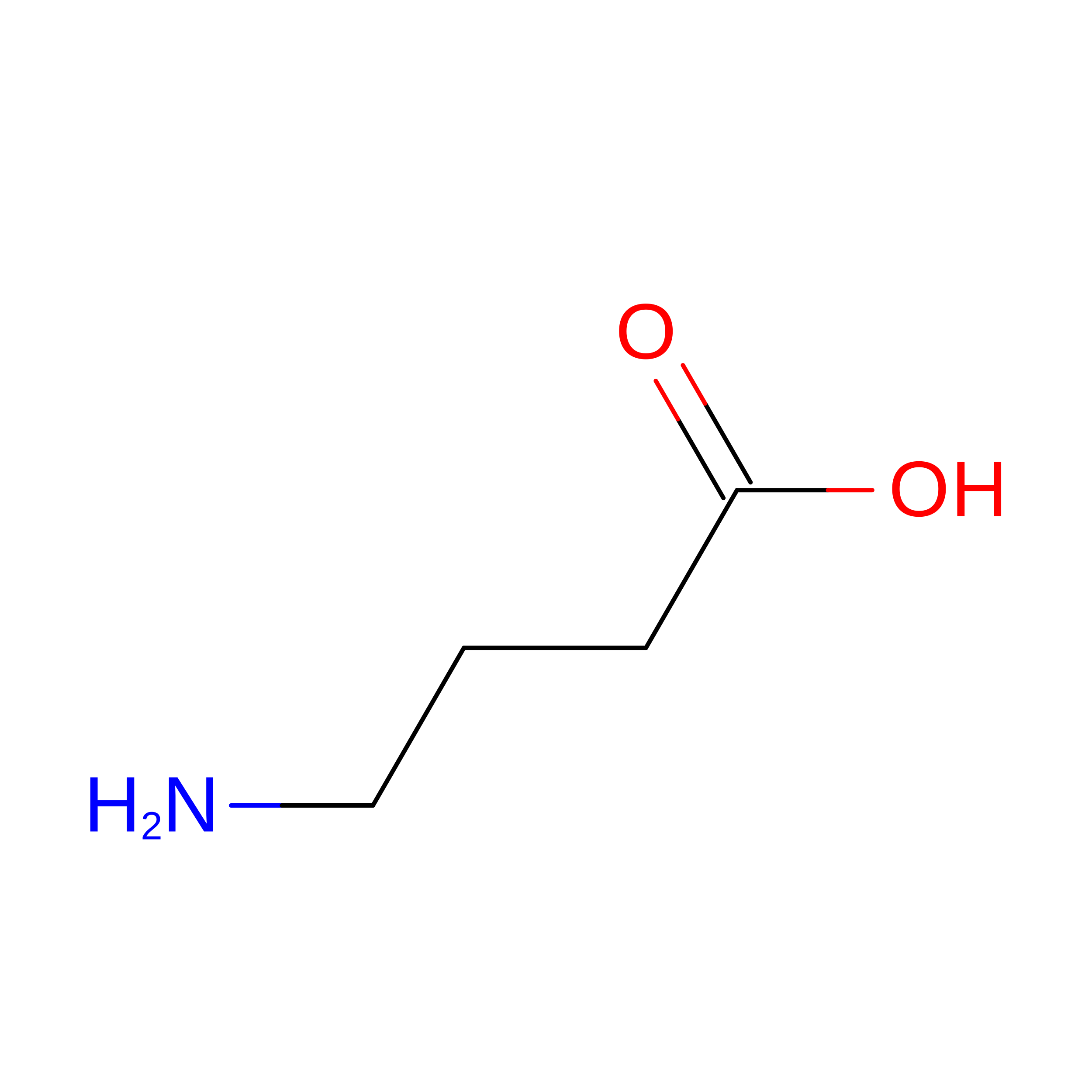
ABU BindingDB:  9OUM Ki: min: 21.37, max: 77.62 (nM) from 4 assay(s)
EC50: min: 230, max: 8.00e+4 (nM) from 20 assay(s)

Experimental Data & Validation

Experimental Data

  • Method: ELECTRON MICROSCOPY
  • Resolution: 2.90 Å
  • Aggregation State: PARTICLE 
  • Reconstruction Method: SINGLE PARTICLE 

Structure Validation

View Full Validation Report


Entry History 

& Funding Information

Deposition Data


Funding OrganizationLocationGrant Number
Howard Hughes Medical Institute (HHMI)United States--

Revision History  (Full details and data files)

  • Version 1.0: 2026-02-18
    Type: Initial release