9OP1 | pdb_00009op1

Cryo-EM structure of Candida albicans fluoride channel FEX in complex with Fab fragment


Experimental Data Snapshot

  • Method: ELECTRON MICROSCOPY
  • Resolution: 4.05 Å
  • Aggregation State: PARTICLE 
  • Reconstruction Method: SINGLE PARTICLE 

Starting Models: in silico, experimental
View more details

wwPDB Validation 3D Report Full Report

Validation slider image for 9OP1

This is version 1.2 of the entry. See complete history

Literature

The molecular mechanism of fluoride export by the eukaryotic fluoride channel FEX.

Kang, C.Y.An, M.Heidari, S.Torabifard, H.Ohi, M.D.Stockbridge, R.B.

(2025) Nat Commun 17: 589-589

  • DOI: https://doi.org/10.1038/s41467-025-67289-0
  • Primary Citation Related Structures: 
    9OP1

  • PubMed Abstract: 

    Much of life on Earth, including plants, fungi, and bacteria, evolved to resist toxic environmental fluoride. In eukaryotes, the major resistance mechanism is fluoride export by membrane proteins known as FEX. Using electrophysiology and transport assays, we establish that FEX from plants and yeasts are highly selective fluoride channels. Fluoride transport activity depends on reversible sodium ion binding, but sodium itself is not transported. We determine a structure of a FEX protein, from pathogenic yeast Candida albicans, using cryo-EM. Bolstered by mutagenesis studies, this structure reveals a fluoride permeation route through a single phenylalanine-lined pore. Molecular dynamics simulations demonstrate that a cation binding motif adjacent to the pore provides a stable sodium binding site that is accessible from the external aqueous solution. Comparison to the structurally related bacterial fluoride channels, Flucs, provides a glimpse of the evolution of structural and mechanistic complexity in a membrane protein family with inverted repeat architecture.


  • Organizational Affiliation
    • Program in Biophysics, University of Michigan, Ann Arbor, MI, USA.

Macromolecule Content 

  • Total Structure Weight: 86.32 kDa 
  • Atom Count: 5,843 
  • Modeled Residue Count: 766 
  • Deposited Residue Count: 795 
  • Unique protein chains: 3

Macromolecules

Find similar proteins by:|  3D Structure
Entity ID: 1
MoleculeChains  Sequence LengthOrganismDetailsImage
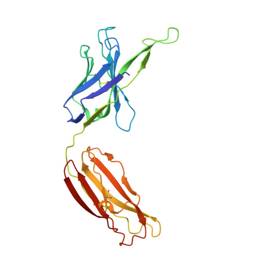
10E8v4 Fab Heavy Chian232Homo sapiensMutation(s): 0 
Find similar proteins by:|  3D Structure
Entity ID: 2
MoleculeChains  Sequence LengthOrganismDetailsImage
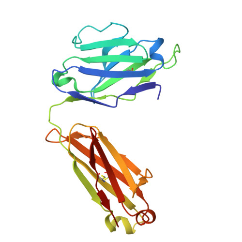
10E8v4 Fab Light Chain210Homo sapiensMutation(s): 0 
Find similar proteins by:|  3D Structure
Entity ID: 3
MoleculeChains  Sequence LengthOrganismDetailsImage
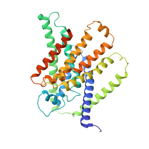
Transmembrane protein gp41,Fluoride export protein 1353HIV-1 M:B_MNCandida albicans SC5314
This entity is chimeric
Mutation(s): 0 
Gene Names: envFEX1CAALFM_C700270WACaO19.7095orf19.7095
UniProt
Find proteins for P05877 (Human immunodeficiency virus type 1 group M subtype B (isolate MN))
Explore P05877 
Go to UniProtKB:  P05877
Find proteins for Q5AFH3 (Candida albicans (strain SC5314 / ATCC MYA-2876))
Explore Q5AFH3 
Go to UniProtKB:  Q5AFH3
Entity Groups
Sequence Clusters30% Identity50% Identity70% Identity90% Identity95% Identity100% Identity
UniProt GroupsP05877Q5AFH3
Sequence Annotations
Expand
Reference Sequence

Experimental Data & Validation

Experimental Data

  • Method: ELECTRON MICROSCOPY
  • Resolution: 4.05 Å
  • Aggregation State: PARTICLE 
  • Reconstruction Method: SINGLE PARTICLE 
EM Software:
TaskSoftware PackageVersion
MODEL REFINEMENTPHENIX1.21_5207
RECONSTRUCTIONcryoSPARC4.5

Structure Validation

View Full Validation Report


Entry History 

& Funding Information

Deposition Data


Funding OrganizationLocationGrant Number
National Institutes of Health/National Institute of General Medical Sciences (NIH/NIGMS)United StatesGM128768

Revision History  (Full details and data files)

  • Version 1.0: 2025-12-03
    Type: Initial release
  • Version 1.1: 2025-12-24
    Changes: Data collection, Database references
  • Version 1.2: 2026-01-28
    Changes: Data collection, Database references