9OLM | pdb_00009olm

Structure of human TRPC3 cerebellar splice variant (isoform c)


Experimental Data Snapshot

  • Method: ELECTRON MICROSCOPY
  • Resolution: 2.50 Å
  • Aggregation State: PARTICLE 
  • Reconstruction Method: SINGLE PARTICLE 

Starting Model: experimental
View more details

wwPDB Validation 3D Report Full Report

Validation slider image for 9OLM

This is version 1.1 of the entry. See complete history

Literature

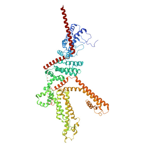
Functional and structural basis of a hypermorphic TRPC3 variant.

Bell, B.Jaramillo-Granada, A.M.Romero, L.O.Gutierrez, I.A.Mallampalli, V.K.P.S.Fan, G.Varma, S.Baker, M.L.Serysheva, I.I.Vasquez, V.Cordero-Morales, J.F.

(2026) Sci Adv 12: eaec9284-eaec9284

  • DOI: https://doi.org/10.1126/sciadv.aec9284
  • Primary Citation Related Structures: 
    9OLK, 9OLL, 9OLM, 9OLX, 9OPU

  • PubMed Abstract: 

    Cerebellar ataxias are characterized by impaired motor coordination resulting from neuronal dysfunction within the cerebellum. The mechanisms underlying this pathology and its cerebellar-specific neurodegeneration remain unknown. We uncover how a gain-of-function canonical transient receptor potential member 3 (TRPC3) mutation, coupled with a cerebellum-specific isoform, stabilizes the channel's open state, resists the leading inhibitor Pyr3, and drives calcium-dependent cell death. Restoring calcium homeostasis by expressing a Purkinje cell calcium pump improves cell viability. Transgenic expression of the TRPC3 hypermorphic variant in Caenorhabditis elegans induces neurodegeneration, confirming its pathogenicity across species. Cryo-electron microscopy and molecular simulations reveal the structural basis for the stabilization of the cerebellar-specific TRPC3 variant in its open state and uncover a druggable allosteric inhibitory binding site. These findings provide an explanation for the vulnerability of cerebellar neurons in TRPC3-associated ataxias and highlight a site for therapeutic intervention.


  • Organizational Affiliation
    • Department of Biochemistry and Molecular Biology, Center for Membrane Biology, McGovern Medical School at the University of Texas Health Science Center at Houston, Houston, TX, USA.

Macromolecule Content 

  • Total Structure Weight: 376.73 kDa 
  • Atom Count: 24,528 
  • Modeled Residue Count: 2,980 
  • Deposited Residue Count: 3,280 
  • Unique protein chains: 1

Macromolecules

Find similar proteins by:|  3D Structure
Entity ID: 1
MoleculeChains  Sequence LengthOrganismDetailsImage
Short transient receptor potential channel 3
A, B, C, D
820Homo sapiensMutation(s): 0 
Gene Names: TRPC3TRP3
UniProt & NIH Common Fund Data Resources
Find proteins for Q13507 (Homo sapiens)
Explore Q13507 
Go to UniProtKB:  Q13507
PHAROS:  Q13507
GTEx:  ENSG00000138741 
Entity Groups
Sequence Clusters30% Identity50% Identity70% Identity90% Identity95% Identity100% Identity
UniProt GroupQ13507
Sequence Annotations
Expand
Reference Sequence

Experimental Data & Validation

Experimental Data

  • Method: ELECTRON MICROSCOPY
  • Resolution: 2.50 Å
  • Aggregation State: PARTICLE 
  • Reconstruction Method: SINGLE PARTICLE 
EM Software:
TaskSoftware PackageVersion
MODEL REFINEMENTPHENIX
RECONSTRUCTIONcryoSPARC

Structure Validation

View Full Validation Report


Entry History 

& Funding Information

Deposition Data


Funding OrganizationLocationGrant Number
National Institutes of Health/National Institute of General Medical Sciences (NIH/NIGMS)United StatesR35GM149218

Revision History  (Full details and data files)

  • Version 1.0: 2026-03-25
    Type: Initial release
  • Version 1.1: 2026-04-08
    Changes: Data collection, Database references