9O4X | pdb_00009o4x

Gamma delta T cell receptor bound to CD1d


Experimental Data Snapshot

  • Method: X-RAY DIFFRACTION
  • Resolution: 2.86 Å
  • R-Value Free: 
    0.255 (Depositor), 0.259 (DCC) 
  • R-Value Work: 
    0.215 (Depositor), 0.220 (DCC) 
  • R-Value Observed: 
    0.218 (Depositor) 

Starting Model: experimental
View more details

wwPDB Validation 3D Report Full Report

Validation slider image for 9O4X

Ligand Structure Quality Assessment 


This is version 1.3 of the entry. See complete history

Literature

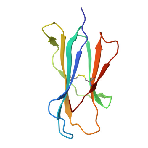
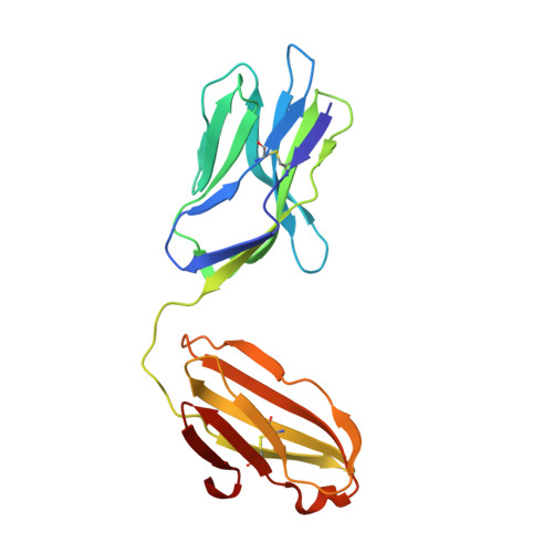
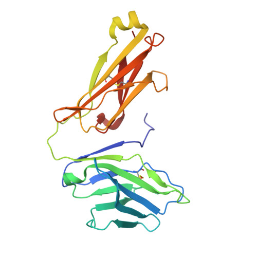
gamma delta T cell receptor recognition of CD1d in a lipid-independent manner.

Rice, M.T.Gunasinghe, S.D.Sok, C.L.Pan, M.Lay, C.S.Gully, B.S.Rossjohn, J.

(2025) Nat Commun 17: 926-926

  • DOI: https://doi.org/10.1038/s41467-025-67653-0
  • Primary Citation Related Structures: 
    9O4X

  • PubMed Abstract: 

    The monomorphic antigen-presenting molecule CD1d presents lipid antigens to both αβ and γδ T cells. While type I natural killer T cells (NKT) display exquisite specificity for CD1d presenting α-galactosylceramide (α-GalCer), the extent of lipid specificity exhibited by CD1d-restricted γδ T cells remains unclear. Here, we demonstrate that human γδ T cell receptors (TCRs) can recognise CD1d in either a lipid-dependent or lipid-independent manner with weak to moderate affinity. Using small-angle X-Ray scattering, we find that γδ TCR-CD1d binding modality is conserved across distinct CD1d-restricted TCRs. In functional assays, CD1d γδ TCR affinity was a poor predictor of γδ T cell line activation. Moreover, CD1d presenting endogenous lipids was sufficient to stimulate T cell activation and induce γδ TCR-CD3 clustering and phosphorylation in a dose-dependent manner. Elongation of the γδ TCR-CD3 complex by the inclusion of the Cγ2 and Cγ3 -encoded constant domains perturbed cellular activation whilst maintaining the ability to form functional γδ TCR clusters. The crystal structure of a Vδ1 γδ + TCR-CD1d complex showed that the γδ TCR sat atop of the CD1d antigen-binding cleft but made no contacts with the presented lipid antigen. These findings provide a molecular basis for lipid-independent CD1d recognition by γδ TCRs.


  • Organizational Affiliation
    • Infection and Immunity Program and Department of Biochemistry and Molecular Biology, Biomedicine Discovery Institute, Monash University, Clayton, Victoria, Australia.

Macromolecule Content 

  • Total Structure Weight: 92.98 kDa 
  • Atom Count: 6,542 
  • Modeled Residue Count: 794 
  • Deposited Residue Count: 794 
  • Unique protein chains: 4

Macromolecules

Find similar proteins by:|  3D Structure
Entity ID: 1
MoleculeChains  Sequence LengthOrganismDetailsImage
Beta-2-microglobulinA [auth B]99Homo sapiensMutation(s): 0 
Gene Names: B2MCDABP0092HDCMA22P
UniProt & NIH Common Fund Data Resources
Find proteins for P61769 (Homo sapiens)
Explore P61769 
Go to UniProtKB:  P61769
PHAROS:  P61769
GTEx:  ENSG00000166710 
Entity Groups
Sequence Clusters30% Identity50% Identity70% Identity90% Identity95% Identity100% Identity
UniProt GroupP61769
Sequence Annotations
Expand
Reference Sequence
Find similar proteins by:|  3D Structure
Entity ID: 2
MoleculeChains  Sequence LengthOrganismDetailsImage
T cell receptor delta chainB [auth D]197Homo sapiensMutation(s): 0 
Find similar proteins by:|  3D Structure
Entity ID: 3
MoleculeChains  Sequence LengthOrganismDetailsImage
T cell receptor gamma chainC [auth G]224Homo sapiensMutation(s): 0 
Find similar proteins by:|  3D Structure
Entity ID: 4
MoleculeChains  Sequence LengthOrganismDetailsImage
Antigen-presenting glycoprotein CD1dD [auth A]274Homo sapiensMutation(s): 0 
Gene Names: CD1D
UniProt & NIH Common Fund Data Resources
Find proteins for P15813 (Homo sapiens)
Explore P15813 
Go to UniProtKB:  P15813
PHAROS:  P15813
GTEx:  ENSG00000158473 
Entity Groups
Sequence Clusters30% Identity50% Identity70% Identity90% Identity95% Identity100% Identity
UniProt GroupP15813
Glycosylation
Glycosylation Sites: 1Go to GlyGen: P15813-1
Sequence Annotations
Expand
Reference Sequence

Oligosaccharides

Help  
Entity ID: 5
MoleculeChains Length2D Diagram GlycosylationD Interactions
2-acetamido-2-deoxy-beta-D-glucopyranose-(1-4)-2-acetamido-2-deoxy-beta-D-glucopyranoseE [auth C],
F [auth E]
2N-Glycosylation
Glycosylation Resources
GlyTouCan: G42666HT
GlyCosmos: G42666HT
GlyGen: G42666HT

Experimental Data & Validation

Experimental Data

  • Method: X-RAY DIFFRACTION
  • Resolution: 2.86 Å
  • R-Value Free:  0.255 (Depositor), 0.259 (DCC) 
  • R-Value Work:  0.215 (Depositor), 0.220 (DCC) 
  • R-Value Observed: 0.218 (Depositor) 
Space Group: P 43 21 2
Unit Cell:
Length ( Å )Angle ( ˚ )
a = 178.093α = 90
b = 178.093β = 90
c = 75.428γ = 90
Software Package:
Software NamePurpose
PHENIXrefinement
XDSdata reduction
XDSdata scaling
PHENIXphasing

Structure Validation

View Full Validation Report


Ligand Structure Quality Assessment 


Entry History 

& Funding Information

Deposition Data


Funding OrganizationLocationGrant Number
Australian Research Council (ARC)Australia--

Revision History  (Full details and data files)

  • Version 1.0: 2025-12-10
    Type: Initial release
  • Version 1.1: 2026-01-14
    Changes: Database references
  • Version 1.2: 2026-01-21
    Changes: Database references
  • Version 1.3: 2026-02-04
    Changes: Database references