9O1K | pdb_00009o1k

Pseudomonas aeruginosa ATPase State3 Fo focused


Experimental Data Snapshot

  • Method: ELECTRON MICROSCOPY
  • Resolution: 3.01 Å
  • Aggregation State: PARTICLE 
  • Reconstruction Method: SINGLE PARTICLE 

wwPDB Validation 3D Report Full Report

Validation slider image for 9O1K

This is version 1.2 of the entry. See complete history

Literature

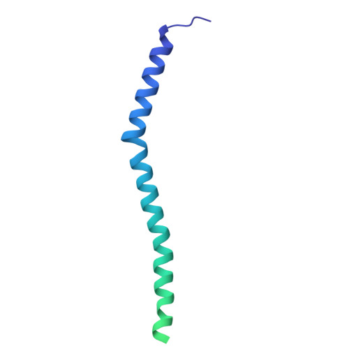
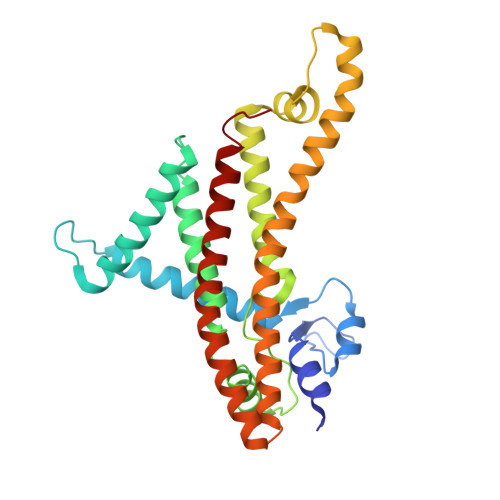
Distinct structural features of Pseudomonas aeruginosa ATP synthase revealed by cryo-electron microscopy.

Sobti, M.Gunn, A.P.Brown, S.H.J.Zavan, L.Fraunfelter, V.M.Wolfe, A.L.McDevitt, C.A.Steed, P.R.Stewart, A.G.

(2025) Nat Commun 17: 406-406

  • DOI: https://doi.org/10.1038/s41467-025-67100-0
  • Primary Citation Related Structures: 
    9O19, 9O1A, 9O1B, 9O1C, 9O1D, 9O1E, 9O1F, 9O1G, 9O1H, 9O1J, 9O1K

  • PubMed Abstract: 

    F 1 F o ATP synthase is the ubiquitous enzyme that synthesizes cellular ATP by coupling proton-motive force with rotational catalysis. Structural differences between prokaryotic and eukaryotic ATP synthases offer potential targets for antimicrobial development. Here, we present the 2.0-2.4 Å resolution cryo-electron microscopy structures of the ATP synthase from Pseudomonas aeruginosa, an opportunistic bacterial pathogen capable of causing serious infections in humans. Our structures identify two distinctive features of this species' enzyme: a distinct binding site for the inhibitory ε subunit, and a coordinated metal ion capping the cytoplasmic proton channel. Lower-resolution maps of the enzyme following incubation with MgATP showed conformational rearrangements of the ε subunit during activation. Visualization of bound water molecules in the periplasmic half-channel supports a Grotthuss proton-transfer mechanism. Focused classification of the F o motor resolves distinct ~11° sub-steps in the c-ring, corresponding to protonation and deprotonation events. Functional analyses show that modifications to either the ε subunit or the metal binding site influence ATP synthesis and hydrolysis. Mass spectrometry analyses suggests that the physiological metal within the complex is zinc. Collectively, these findings define structural features of P. aeruginosa ATP synthase that could serve as targets for antimicrobial therapeutics.


  • Organizational Affiliation
    • Molecular, Structural and Computational Biology Division, The Victor Chang Cardiac Research Institute, Darlinghurst, NSW, Australia.

Macromolecule Content 

  • Total Structure Weight: 152.02 kDa 
  • Atom Count: 9,090 
  • Modeled Residue Count: 1,232 
  • Deposited Residue Count: 1,451 
  • Unique protein chains: 3

Macromolecules

Find similar proteins by:|  3D Structure
Entity ID: 1
MoleculeChains  Sequence LengthOrganismDetailsImage
ATP synthase subunit c85Pseudomonas aeruginosaMutation(s): 0 
Gene Names: atpEPSPA7_6361
UniProt
Find proteins for A6VF37 (Pseudomonas paraeruginosa (strain DSM 24068 / PA7))
Explore A6VF37 
Go to UniProtKB:  A6VF37
Entity Groups
Sequence Clusters30% Identity50% Identity70% Identity90% Identity95% Identity100% Identity
UniProt GroupA6VF37
Sequence Annotations
Expand
Reference Sequence
Find similar proteins by:|  3D Structure
Entity ID: 2
MoleculeChains  Sequence LengthOrganismDetailsImage
ATP synthase subunit bK [auth X],
L [auth Y]
156Pseudomonas aeruginosaMutation(s): 0 
Gene Names: atpFPA14_73290
UniProt
Find proteins for A6VF36 (Pseudomonas paraeruginosa (strain DSM 24068 / PA7))
Explore A6VF36 
Go to UniProtKB:  A6VF36
Entity Groups
Sequence Clusters30% Identity50% Identity70% Identity90% Identity95% Identity100% Identity
UniProt GroupA6VF36
Sequence Annotations
Expand
Reference Sequence
Find similar proteins by:|  3D Structure
Entity ID: 3
MoleculeChains  Sequence LengthOrganismDetailsImage
ATP synthase subunit aM [auth a]289Pseudomonas aeruginosaMutation(s): 0 
Gene Names: atpBPSPA7_6362
UniProt
Find proteins for A6VF38 (Pseudomonas paraeruginosa (strain DSM 24068 / PA7))
Explore A6VF38 
Go to UniProtKB:  A6VF38
Entity Groups
Sequence Clusters30% Identity50% Identity70% Identity90% Identity95% Identity100% Identity
UniProt GroupA6VF38
Sequence Annotations
Expand
Reference Sequence

Experimental Data & Validation

Experimental Data

  • Method: ELECTRON MICROSCOPY
  • Resolution: 3.01 Å
  • Aggregation State: PARTICLE 
  • Reconstruction Method: SINGLE PARTICLE 

Structure Validation

View Full Validation Report


Entry History 

& Funding Information

Deposition Data


Funding OrganizationLocationGrant Number
National Health and Medical Research Council (NHMRC, Australia)Australia--

Revision History  (Full details and data files)

  • Version 1.0: 2025-12-10
    Type: Initial release
  • Version 1.1: 2025-12-24
    Changes: Data collection, Database references
  • Version 1.2: 2026-01-28
    Changes: Data collection, Database references