9O0B | pdb_00009o0b

X-ray Crystal Structure of Fission Yeast Fsc1 protein in P43212


Experimental Data Snapshot

  • Method: X-RAY DIFFRACTION
  • Resolution: 2.50 Å
  • R-Value Free: 
    0.298 (Depositor), 0.295 (DCC) 
  • R-Value Work: 
    0.244 (Depositor), 0.240 (DCC) 

wwPDB Validation 3D Report Full Report

Validation slider image for 9O0B

This is version 1.2 of the entry. See complete history

Literature

Crystal structures of Fsc1, a novel autophagy factor that mediates autophagosome-vacuole fusion in fission yeast.

Azuka, C.Liu, J.Jin, X.

(2026) Acta Crystallogr D Struct Biol 82: 358-369

  • DOI: https://doi.org/10.1107/S205979832600197X
  • Primary Citation Related Structures: 
    9NU9, 9O0B

  • PubMed Abstract: 

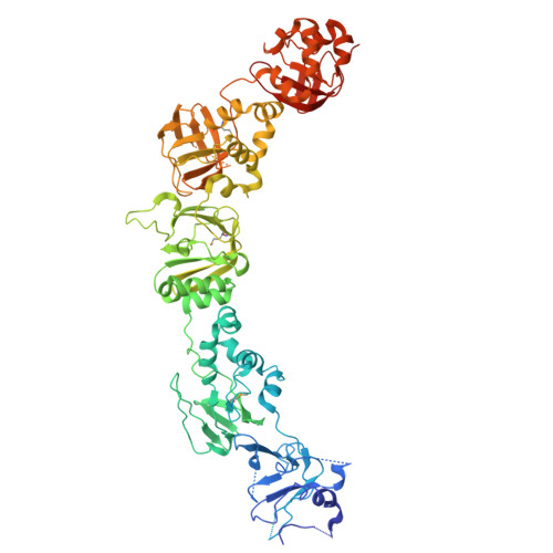
    Fsc1 is a recently identified autophagy factor in the fission yeast Schizosaccharomyces pombe that is implicated in the autophagosome-vacuole fusion step during the final stages of autophagy. Despite its critical role, the structural basis of Fsc1 function has remained unknown. Here, we report the first crystal structures of the luminal domain of Fsc1, revealing an elongated, modular architecture composed of five tandem fasciclin (FAS1) domains. Each domain adopts a hallmark β-sandwich fold, and the overall assembly forms a continuous scaffold featuring a conserved surface groove within the FAS1-4 domain. Structural and biochemical analyses demonstrate that Fsc1 forms a homodimer in solution through a shared interface observed in two independent crystal forms, supporting a biologically relevant but potentially low-affinity association. Comparative sequence and structural analyses reveal significant homology between Fsc1 and human fasciclin proteins, including TGFBI and periostin, suggesting similar structural principles underlying their functions. Together, these findings provide the first structural insights into Fsc1 and establish a structural framework for understanding how its modular architecture and context-dependent dimerization may facilitate late-stage membrane fusion during autophagy.


  • Organizational Affiliation
    • Department of Chemistry, Michigan State University, East Lansing, MI 48824, USA.

Macromolecule Content 

  • Total Structure Weight: 73.79 kDa 
  • Atom Count: 5,002 
  • Modeled Residue Count: 609 
  • Deposited Residue Count: 649 
  • Unique protein chains: 1

Macromolecules

Find similar proteins by:|  3D Structure
Entity ID: 1
MoleculeChains  Sequence LengthOrganismDetailsImage
FAS1 domain-containing protein fsc1649Schizosaccharomyces pombeMutation(s): 0 
Gene Names: fsc1SPAC22H12.05c
UniProt
Find proteins for O94439 (Schizosaccharomyces pombe (strain 972 / ATCC 24843))
Explore O94439 
Go to UniProtKB:  O94439
Entity Groups
Sequence Clusters30% Identity50% Identity70% Identity90% Identity95% Identity100% Identity
UniProt GroupO94439
Sequence Annotations
Expand
Reference Sequence

Small Molecules

Modified Residues  1 Unique
IDChains TypeFormula2D DiagramParent
MSE
Query on MSE
A
L-PEPTIDE LINKINGC5 H11 N O2 SeMET

Experimental Data & Validation

Experimental Data

  • Method: X-RAY DIFFRACTION
  • Resolution: 2.50 Å
  • R-Value Free:  0.298 (Depositor), 0.295 (DCC) 
  • R-Value Work:  0.244 (Depositor), 0.240 (DCC) 
Space Group: P 43 21 2
Unit Cell:
Length ( Å )Angle ( ˚ )
a = 58.003α = 90
b = 58.003β = 90
c = 488.271γ = 90
Software Package:
Software NamePurpose
REFMACrefinement
HKL-2000data reduction
HKL-2000data scaling
PHENIXphasing

Structure Validation

View Full Validation Report


Entry History 

& Funding Information

Deposition Data


Funding OrganizationLocationGrant Number
Other private--

Revision History  (Full details and data files)

  • Version 1.0: 2026-04-01
    Type: Initial release
  • Version 1.1: 2026-04-08
    Changes: Database references
  • Version 1.2: 2026-04-15
    Changes: Database references