9O01 | pdb_00009o01

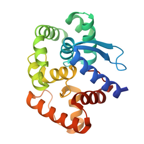
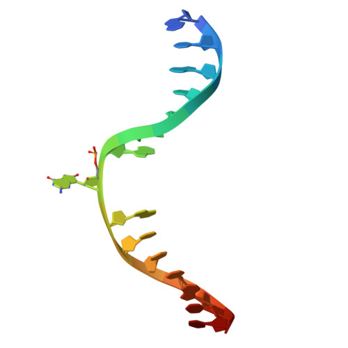
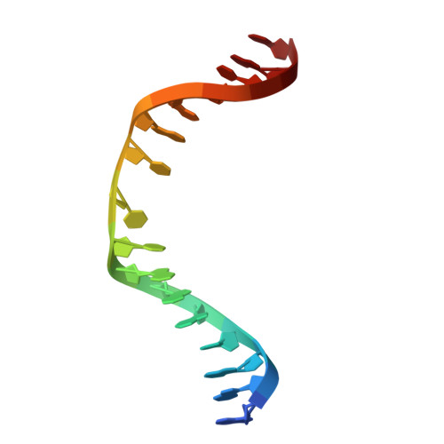
Crystal structure of AfOgg1-D140N mutant bound to 8-OG DNA duplex in an intermediate state


Experimental Data Snapshot

  • Method: X-RAY DIFFRACTION
  • Resolution: 1.95 Å
  • R-Value Free: 
    0.260 (Depositor), 0.260 (DCC) 
  • R-Value Work: 
    0.221 (Depositor), 0.222 (DCC) 
  • R-Value Observed: 
    0.225 (Depositor) 

Starting Model: experimental
View more details

wwPDB Validation 3D Report Full Report

Validation slider image for 9O01

This is version 1.1 of the entry. See complete history

Literature

A unified catalytic mechanism in bifunctional DNA glycosylases with an evolutionarily conserved aspartate-lysine dyad.

Syed, A.Serafim, L.F.Arvai, A.S.Minko, I.G.Tang, H.Y.H.Huffman, J.L.Mol, C.D.Hitomi, K.Sarker, A.H.Parikh, S.Tsai, C.L.Bacolla, A.Shin, D.S.Cunningham, R.P.Iwai, S.Chowdhury, D.Lloyd, R.S.Ivanov, I.Tainer, J.A.

(2026) Nat Commun 

  • DOI: https://doi.org/10.1038/s41467-026-75471-1
  • Primary Citation Related Structures: 
    10MN, 9NZ8, 9NZ9, 9NZA, 9NZC, 9NZD, 9O00, 9O01, 9O02, 9O03

  • PubMed Abstract: 

    Bifunctional glycosylases, OGG1 for purines and NTH1 for pyrimidines, repair oxidized DNA bases via consecutive glycosylase and AP-lyase reactions, yet their catalytic relationships and lyase activity's biological relevance remain unresolved. Here, we solved crystal structures of archaeal and human Ogg1 and Nth1 captured in key damage-recognition and catalysis-ready states, complemented by ab initio molecular dynamics simulations of their complete reaction trajectories. We thereby define a unified catalytic mechanism for OGG1 and NTH1 conserved over three billion years, distinct from the canonical oxocarbenium-ion mechanism of monofunctional glycosylases. While divergent in their oxidized substrate recognition, their chemistry converged on ribose protonation and ring opening that precede the deglycosylation step. Acid-base catalysis mediated by a conserved aspartate-lysine dyad lowers the C-N bond cleavage barrier, while the excised 8-oxoG base in OGG1 or a conserved aspartate in NTH1 facilitates the AP-lyase reaction. Moreover, structures of human OGG1 bound to product DNA and to product DNA plus a potent small-molecule agonist F51, within the catalytic pocket, reveal that agonists accelerate enzyme turnover by promoting product release. Together, these findings clarify the catalytic logic of bifunctional glycosylases, enabling the development of chemical tools to interrogate lyase activity and therapeutics for oxidative damage in cancer and aging.


  • Organizational Affiliation
    • Department of Molecular and Cellular Oncology, Department of Cancer Biology, The University of Texas MD Anderson Cancer Center, Houston, TX, USA. aleem_syed@dfci.harvard.edu.

Macromolecule Content 

  • Total Structure Weight: 33.01 kDa 
  • Atom Count: 2,288 
  • Modeled Residue Count: 229 
  • Deposited Residue Count: 235 
  • Unique protein chains: 1
  • Unique nucleic acid chains: 2

Macromolecules


Find similar proteins by:|  3D Structure
Entity ID: 1
MoleculeChains  Sequence LengthOrganismDetailsImage
8-oxoguanine DNA glycosylase/AP lyase203Archaeoglobus fulgidusMutation(s): 1 
Gene Names: oggAF_0371
EC: 3.2.2 (PDB Primary Data), 4.2.99.18 (PDB Primary Data)
UniProt
Find proteins for O29876 (Archaeoglobus fulgidus (strain ATCC 49558 / DSM 4304 / JCM 9628 / NBRC 100126 / VC-16))
Explore O29876 
Go to UniProtKB:  O29876
Entity Groups
Sequence Clusters30% Identity50% Identity70% Identity90% Identity95% Identity100% Identity
UniProt GroupO29876
Sequence Annotations
Expand
Reference Sequence
Find similar nucleic acids by:  Sequence
Entity ID: 2
MoleculeChains LengthOrganismImage
DNA (5'-D(P*GP*CP*GP*TP*CP*CP*AP*(8OG)P*GP*TP*CP*TP*AP*CP*CP*T)-3')16synthetic construct
Sequence Annotations
Expand
Reference Sequence
Find similar nucleic acids by:  Sequence
Entity ID: 3
MoleculeChains LengthOrganismImage
DNA (5'-D(*AP*GP*GP*TP*AP*GP*AP*CP*CP*TP*GP*GP*AP*CP*GP*C)-3')16synthetic construct
Sequence Annotations
Expand
Reference Sequence

Small Molecules

Ligands 1 Unique
IDChains Name / Formula / InChI Key2D Diagram3D Interactions
MG

Query on MG


Download:Ideal Coordinates CCD File
D [auth A]MAGNESIUM ION
Mg
JLVVSXFLKOJNIY-UHFFFAOYSA-N

Experimental Data & Validation

Experimental Data

  • Method: X-RAY DIFFRACTION
  • Resolution: 1.95 Å
  • R-Value Free:  0.260 (Depositor), 0.260 (DCC) 
  • R-Value Work:  0.221 (Depositor), 0.222 (DCC) 
  • R-Value Observed: 0.225 (Depositor) 
Space Group: P 1 21 1
Unit Cell:
Length ( Å )Angle ( ˚ )
a = 52.013α = 90
b = 52.839β = 100.46
c = 53.459γ = 90
Software Package:
Software NamePurpose
PHENIXrefinement
XDSdata reduction
XDSdata scaling
PHASERphasing

Structure Validation

View Full Validation Report


Entry History 

& Funding Information

Deposition Data


Funding OrganizationLocationGrant Number
National Institutes of Health/National Cancer Institute (NIH/NCI)United StatesP01 CA092584
National Institutes of Health/National Cancer Institute (NIH/NCI)United StatesR35 CA220430

Revision History  (Full details and data files)

  • Version 1.0: 2026-04-15
    Type: Initial release
  • Version 1.1: 2026-08-05
    Changes: Database references