9NY8 | pdb_00009ny8

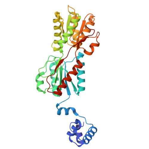
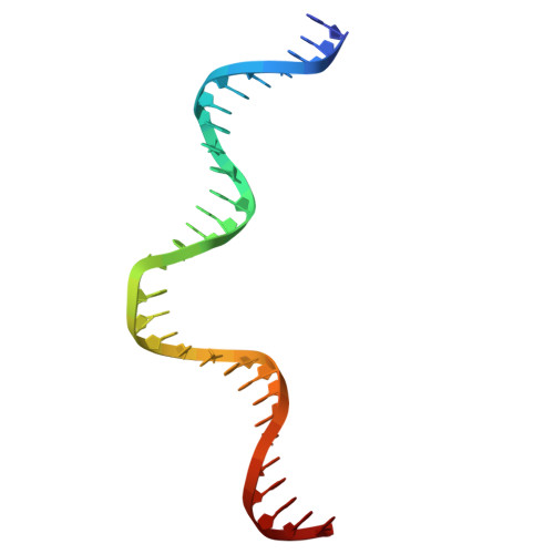
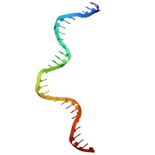
Crystal structure of the ribose operon repressor, RbsR, bound to ribose operon


Experimental Data Snapshot

  • Method: X-RAY DIFFRACTION
  • Resolution: 2.10 Å
  • R-Value Free: 
    0.223 (Depositor), 0.202 (DCC) 
  • R-Value Work: 
    0.192 (Depositor) 
  • R-Value Observed: 
    0.194 (Depositor) 

Starting Model: in silico
View more details

wwPDB Validation 3D Report Full Report

Validation slider image for 9NY8

Ligand Structure Quality Assessment 


This is version 1.1 of the entry. See complete history. 

Literature

Distinct energetic blueprints diversify function of conserved protein folds.

Wells, M.L., Lu, C., Sultanov, D., Weber, K.C., Ahern, E., Gong, Z., Chen, E., Glasgow, A.

(2026) Nat Chem 18: 1496-1506

  • DOI: https://doi.org/10.1038/s41557-026-02163-0
  • Primary Citation Related Structures: 
    9NY7, 9NY8

  • PubMed Abstract: 

    Advances in structural biology have improved our understanding of the relationship between protein structure and function, while also confirming a widely applicable principle: protein domains with highly conserved three-dimensional folds can perform radically disparate biochemical functions. To gain insight into this structural enigma, we mapped the energetic landscapes of a family of bacterial transcription factors and their anciently diverged structural homologues, the periplasmic binding proteins. Using hydrogen exchange-mass spectrometry, bioinformatics, X-ray crystallography and molecular dynamics, we uncovered an unexpected contrast: despite binding the same sugars, the two families have evolved unique 'energetic blueprints' to support their distinct functional requirements. To test if differences in ensemble energies have functional consequences, we rationally redesigned the protein fold for tunable ligand-driven transcriptional responses. Strikingly, energy-driven protein engineering produced synthetic transcription factors with the theoretically anticipated ligand-induced transcriptional outputs. Thus, decoding energetic blueprints among conserved protein folds provides diverse functional adaptations, paves an alternative roadmap for protein design, and offers a distinct approach for engineering challenging drug targets.


  • Organizational Affiliation: 
    • Department of Biochemistry and Molecular Biophysics, Columbia University Irving Medical Center, New York, NY, USA.

Macromolecule Content 

  • Total Structure Weight: 91.95 kDa 
  • Atom Count: 6,439 
  • Modeled Residue Count: 720 
  • Deposited Residue Count: 720 
  • Unique protein chains: 1
  • Unique nucleic acid chains: 2

Macromolecules


Find similar proteins by:|  3D Structure
Entity ID: 1
MoleculeChains  Sequence LengthOrganismDetailsImage
Ribose operon repressor
A, B
330Escherichia coliMutation(s): 0 
Gene Names: rbsR, b3753, JW3732
UniProt
Find proteins for P0ACQ0 (Escherichia coli (strain K12))
Explore P0ACQ0 
Go to UniProtKB:  P0ACQ0
Entity Groups
Sequence Clusters30% Identity50% Identity70% Identity90% Identity95% Identity100% Identity
UniProt GroupP0ACQ0
Sequence Annotations
Expand
Reference Sequence
Find similar nucleic acids by:  (by identity cutoff) 
Entity ID: 2
MoleculeChains LengthOrganismImage
ribose operon30Escherichia coli
Sequence Annotations
Expand
Reference Sequence
Find similar nucleic acids by:  (by identity cutoff) 
Entity ID: 3
MoleculeChains LengthOrganismImage
ribose operon30Escherichia coli
Sequence Annotations
Expand
Reference Sequence

Experimental Data & Validation

Experimental Data

  • Method: X-RAY DIFFRACTION
  • Resolution: 2.10 Å
  • R-Value Free:  0.223 (Depositor), 0.202 (DCC) 
  • R-Value Work:  0.192 (Depositor) 
  • R-Value Observed: 0.194 (Depositor) 
Space Group: P 31 2 1
Unit Cell:
Length ( Å )Angle ( ˚ )
a = 125.53α = 90
b = 125.53β = 90
c = 122.31γ = 120
Software Package:
Software NamePurpose
REFMACrefinement
xia2data reduction
STARANISOdata scaling
Aimlessdata scaling
PHASERphasing

Structure Validation

View Full Validation Report


Ligand Structure Quality Assessment 


Entry History 

& Funding Information

Deposition Data


Funding OrganizationLocationGrant Number
National Institutes of Health/National Institute of General Medical Sciences (NIH/NIGMS)United StatesR00GM135529

Revision History  (Full details and data files)

  • Version 1.0: 2025-07-16
    Type: Initial release
  • Version 1.1: 2026-10-14
    Changes: Database references