9NWB | pdb_00009nwb

Crystal structure of SARS-CoV-2 main protease in complex with an inhibitor TKB-280-5I


Experimental Data Snapshot

  • Method: X-RAY DIFFRACTION
  • Resolution: 2.01 Å
  • R-Value Free: 
    0.248 (Depositor), 0.224 (DCC) 
  • R-Value Work: 
    0.208 (Depositor) 
  • R-Value Observed: 
    0.210 (Depositor) 

Starting Model: experimental
View more details

wwPDB Validation 3D Report Full Report

Validation slider image for 9NWB

Ligand Structure Quality Assessment 


This is version 1.1 of the entry. See complete history

Literature

Impact of Single Halogen Atom Substitutions on Antiviral Profile of Inhibitors Targeting SARS-CoV‐2 Main Protease.

Bulut, H.Higashi-Kuwata, N.Ogata-Aoki, H.Hayashi, H.Takamune, N.Kishimoto, N.Das, D.Li, M.Wlodawer, A.Misumi, S.Tamamura, H.Mitsuya, H.

(2026) ACS Omega 11: 4541-4550

  • DOI: https://doi.org/10.1021/acsomega.5c10895
  • Primary Citation Related Structures: 
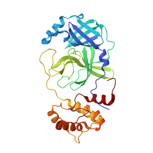
    9NWA, 9NWB, 9NWC

  • PubMed Abstract: 

    The SARS-CoV-2 main protease (M pro ) remains a prime antiviral target because its inhibition halts viral replication. To probe how subtle atomic changes influence drug performance, we carried out a systematic halogen scan on a potent ketoamide scaffold, replacing a single fluorine with chlorine, bromine, or iodine. Enzymatic assays revealed that the F- and Cl-substituted analogues inhibit M pro at nanomolar levels, whereas Br and I variants are 10- to 20-fold weaker. Cell-based antiviral tests mirrored this trend, yet uptake studies showed the opposite: iodine markedly enhances intracellular accumulation. High-resolution X-ray structures (1.6-1.8 Å) explain the dichotomy: small halogens fit snugly in the S1' σ-hole pocket, maximizing hydrogen-bond geometry, while bulkier atoms distort binding but create a lipophilic patch that boosts permeability. These data yield the first fluorine-to-iodine structure-activity map for SARS-CoV-2 M pro inhibitors. These findings highlight the critical role of halogen selection in antiviral inhibitor design.


  • Organizational Affiliation
    • Experimental Retrovirology Section, HIV and AIDS Malignancy Branch, National Cancer Institute, NIH, Bethesda, Maryland 20892, United States.

Macromolecule Content 

  • Total Structure Weight: 34.59 kDa 
  • Atom Count: 2,436 
  • Modeled Residue Count: 306 
  • Deposited Residue Count: 306 
  • Unique protein chains: 1

Macromolecules

Find similar proteins by:|  3D Structure
Entity ID: 1
MoleculeChains  Sequence LengthOrganismDetailsImage
ORF1a polyprotein306Severe acute respiratory syndrome coronavirusMutation(s): 0 
Gene Names: ORF1ab
UniProt
Find proteins for A0A8B1JK87 (Severe acute respiratory syndrome coronavirus 2)
Explore A0A8B1JK87 
Go to UniProtKB:  A0A8B1JK87
Entity Groups
Sequence Clusters30% Identity50% Identity70% Identity90% Identity95% Identity100% Identity
UniProt GroupA0A8B1JK87
Sequence Annotations
Expand
Reference Sequence

Small Molecules

Ligands 1 Unique
IDChains Name / Formula / InChI Key2D Diagram3D Interactions
A1B7A

Query on A1B7A


Download:Ideal Coordinates CCD File
B [auth A](1R,2S,5S)-N-{(1S,2R)-1-hydroxy-1-(5-iodo-1,3-benzothiazol-2-yl)-3-[(3R)-2-oxopyrrolidin-3-yl]propan-2-yl}-6,6-dimethyl-3-[3-methyl-N-(trifluoroacetyl)-L-valyl]-3-azabicyclo[3.1.0]hexane-2-carboxamide
C30 H37 F3 I N5 O5 S
AMWSCQMTJKQAAP-XSQCLQQESA-N

Experimental Data & Validation

Experimental Data

  • Method: X-RAY DIFFRACTION
  • Resolution: 2.01 Å
  • R-Value Free:  0.248 (Depositor), 0.224 (DCC) 
  • R-Value Work:  0.208 (Depositor) 
  • R-Value Observed: 0.210 (Depositor) 
Space Group: P 21 21 2
Unit Cell:
Length ( Å )Angle ( ˚ )
a = 45.412α = 90
b = 64.071β = 90
c = 104.988γ = 90
Software Package:
Software NamePurpose
REFMACrefinement
DIALSdata reduction
SCALEPACKdata scaling
REFMACphasing

Structure Validation

View Full Validation Report


Ligand Structure Quality Assessment 


Entry History 

& Funding Information

Deposition Data


Funding OrganizationLocationGrant Number
National Institutes of Health/National Cancer Institute (NIH/NCI)United States--

Revision History  (Full details and data files)

  • Version 1.0: 2025-06-04
    Type: Initial release
  • Version 1.1: 2026-02-18
    Changes: Database references