9NPY | pdb_00009npy

SARS-CoV-2 nsp1 bound to the Rhinolophus lepidus 40S ribosome (local refinement of the 40S head)


Experimental Data Snapshot

  • Method: ELECTRON MICROSCOPY
  • Resolution: 2.10 Å
  • Aggregation State: PARTICLE 
  • Reconstruction Method: SINGLE PARTICLE 

wwPDB Validation 3D Report Full Report

Validation slider image for 9NPY

This is version 1.1 of the entry. See complete history

Literature

SARS-CoV-2 nsp1 mediates broad inhibition of translation in mammals.

Gen, R.Addetia, A.Asarnow, D.Park, Y.J.Quispe, J.Chan, M.C.Brown, J.T.Lee, J.Campbell, M.G.Lapointe, C.P.Veesler, D.

(2025) Cell Rep 44: 115696-115696

  • DOI: https://doi.org/10.1016/j.celrep.2025.115696
  • Primary Citation Related Structures: 
    9NPX, 9NPY

  • PubMed Abstract: 

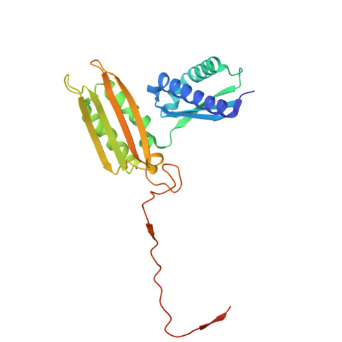
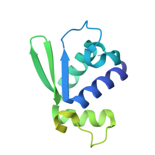
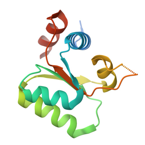
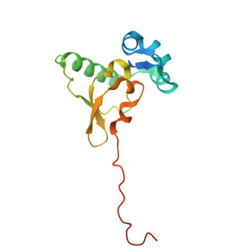
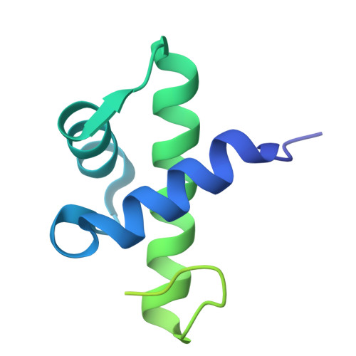
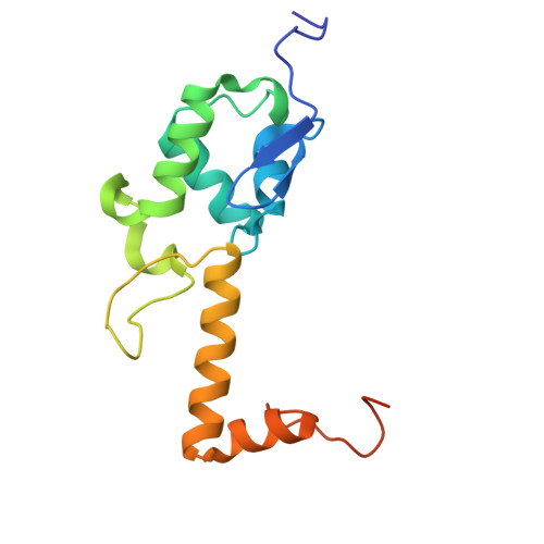
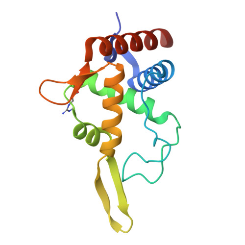
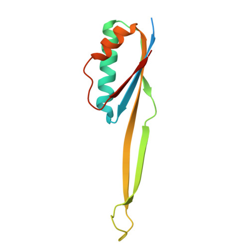
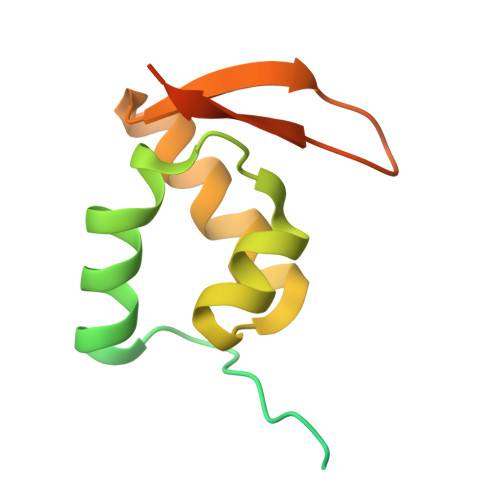
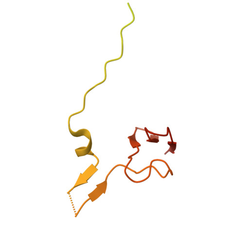
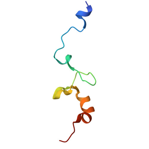
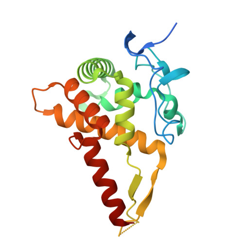
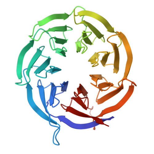
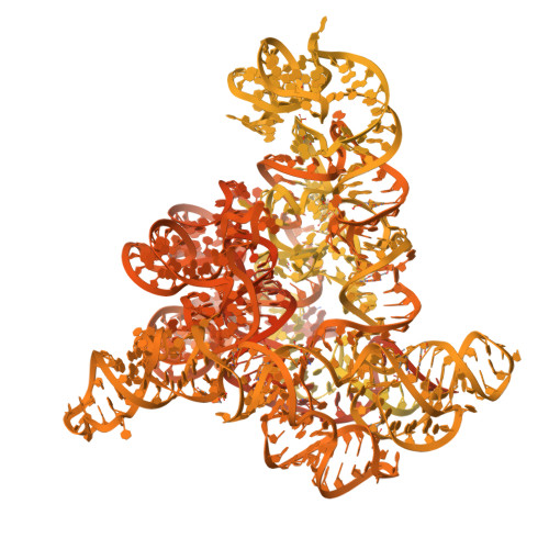
    Severe acute respiratory syndrome coronavirus 2 (SARS-CoV-2) non-structural protein 1 (nsp1) promotes innate immune evasion by inhibiting host translation in human cells. However, the role of nsp1 in other host species remains elusive, especially in bats-natural reservoirs of sarbecoviruses with a markedly different innate immune system than humans. We reveal that nsp1 potently inhibits translation in Rhinolophus lepidus bat cells, which belong to the same genus as known sarbecovirus reservoir hosts. We determined a cryoelectron microscopy structure of nsp1 bound to the R. lepidus 40S ribosomal subunit, showing that it blocks the mRNA entry channel by targeting a highly conserved site among mammals. Accordingly, we found that nsp1 blocked protein translation in mammalian cells from several species, underscoring its broadly inhibitory activity and conserved role in numerous SARS-CoV-2 hosts. Our findings illuminate the arms race between coronaviruses and mammalian host immunity, providing a foundation for understanding the determinants of viral maintenance in bat hosts and spillover.


  • Organizational Affiliation
    • Department of Biochemistry, University of Washington, Seattle, WA 98195, USA.

Macromolecule Content 

  • Total Structure Weight: 865.52 kDa 
  • Atom Count: 25,171 
  • Modeled Residue Count: 2,360 
  • Deposited Residue Count: 4,178 
  • Unique protein chains: 15
  • Unique nucleic acid chains: 1

Macromolecules


Find similar proteins by:|  3D Structure
Entity ID: 1
MoleculeChains  Sequence LengthOrganismDetailsImage
40S ribosomal protein uS3, RPS3A [auth B]243Rhinolophus lepidusMutation(s): 0 
Find similar proteins by:|  3D Structure
Entity ID: 2
MoleculeChains  Sequence LengthOrganismDetailsImage
40S ribosomal protein eS10, RPS10B [auth J]165Rhinolophus lepidusMutation(s): 0 
Find similar proteins by:|  3D Structure
Entity ID: 3
MoleculeChains  Sequence LengthOrganismDetailsImage
40S ribosomal protein eS12, RPS12C [auth L]132Rhinolophus lepidusMutation(s): 0 
Find similar proteins by:|  3D Structure
Entity ID: 4
MoleculeChains  Sequence LengthOrganismDetailsImage
40S ribosomal protein uS19, RPS15D [auth O]145Rhinolophus lepidusMutation(s): 0 
Find similar proteins by:|  3D Structure
Entity ID: 5
MoleculeChains  Sequence LengthOrganismDetailsImage
40S ribosomal protein uS9, RPS16E [auth Q]146Rhinolophus lepidusMutation(s): 0 
Find similar proteins by:|  3D Structure
Entity ID: 6
MoleculeChains  Sequence LengthOrganismDetailsImage
40S ribosomal protein eS17, RPS17F [auth R]135Rhinolophus lepidusMutation(s): 0 
Find similar proteins by:|  3D Structure
Entity ID: 7
MoleculeChains  Sequence LengthOrganismDetailsImage
40S ribosomal protein uS13, RPS18G [auth S]152Rhinolophus lepidusMutation(s): 0 
Find similar proteins by:|  3D Structure
Entity ID: 8
MoleculeChains  Sequence LengthOrganismDetailsImage
40S ribosomal protein eS19, RPS19H [auth T]145Rhinolophus lepidusMutation(s): 0 
Find similar proteins by:|  3D Structure
Entity ID: 9
MoleculeChains  Sequence LengthOrganismDetailsImage
40S ribosomal protein uS10, RPS20I [auth U]119Rhinolophus lepidusMutation(s): 0 
Find similar proteins by:|  3D Structure
Entity ID: 10
MoleculeChains  Sequence LengthOrganismDetailsImage
40S ribosomal protein eS25, RPS25J [auth Y]125Rhinolophus lepidusMutation(s): 0 
Find similar proteins by:|  3D Structure
Entity ID: 11
MoleculeChains  Sequence LengthOrganismDetailsImage
40S ribosomal protein eS31, RPS27aK [auth b]156Rhinolophus lepidusMutation(s): 0 
Find similar proteins by:|  3D Structure
Entity ID: 12
MoleculeChains  Sequence LengthOrganismDetailsImage
40S ribosomal protein eS28, RPS28L [auth c]69Rhinolophus lepidusMutation(s): 0 
Find similar proteins by:|  3D Structure
Entity ID: 13
MoleculeChains  Sequence LengthOrganismDetailsImage
40S ribosomal protein uS14, RPS29M [auth d]56Rhinolophus lepidusMutation(s): 0 
Find similar proteins by:|  3D Structure
Entity ID: 15
MoleculeChains  Sequence LengthOrganismDetailsImage
40S ribosomal protein uS7, RPS5O [auth E]204Rhinolophus lepidusMutation(s): 0 
Find similar proteins by:|  3D Structure
Entity ID: 16
MoleculeChains  Sequence LengthOrganismDetailsImage
RACK1P [auth g]317Rhinolophus lepidusMutation(s): 0 
Find similar nucleic acids by:  (by identity cutoff) 
Entity ID: 14
MoleculeChains LengthOrganismImage
18S ribosomal RNAN [auth i]1,869Rhinolophus lepidus
Sequence Annotations
Expand
Reference Sequence

Small Molecules

Ligands 3 Unique
IDChains Name / Formula / InChI Key2D Diagram3D Interactions
ZN

Query on ZN


Download:Ideal Coordinates CCD File
S [auth b],
T [auth d]
ZINC ION
Zn
PTFCDOFLOPIGGS-UHFFFAOYSA-N
K

Query on K


Download:Ideal Coordinates CCD File
OA [auth i]
PA [auth i]
QA [auth i]
RA [auth i]
SA [auth i]
OA [auth i],
PA [auth i],
QA [auth i],
RA [auth i],
SA [auth i],
TA [auth i],
U [auth d],
UA [auth i],
VA [auth E]
POTASSIUM ION
K
NPYPAHLBTDXSSS-UHFFFAOYSA-N
MG

Query on MG


Download:Ideal Coordinates CCD File
AA [auth i]
BA [auth i]
CA [auth i]
DA [auth i]
EA [auth i]
AA [auth i],
BA [auth i],
CA [auth i],
DA [auth i],
EA [auth i],
FA [auth i],
GA [auth i],
HA [auth i],
IA [auth i],
JA [auth i],
KA [auth i],
LA [auth i],
MA [auth i],
NA [auth i],
Q [auth S],
R [auth T],
V [auth i],
W [auth i],
X [auth i],
Y [auth i],
Z [auth i]
MAGNESIUM ION
Mg
JLVVSXFLKOJNIY-UHFFFAOYSA-N
Modified Residues  1 Unique
IDChains TypeFormula2D DiagramParent
NMM
Query on NMM
H [auth T]L-PEPTIDE LINKINGC7 H16 N4 O2ARG

Experimental Data & Validation

Experimental Data

  • Method: ELECTRON MICROSCOPY
  • Resolution: 2.10 Å
  • Aggregation State: PARTICLE 
  • Reconstruction Method: SINGLE PARTICLE 
EM Software:
TaskSoftware PackageVersion
MODEL REFINEMENTPHENIX1.21.1_5286

Structure Validation

View Full Validation Report


Entry History 

& Funding Information

Deposition Data


Funding OrganizationLocationGrant Number
Howard Hughes Medical Institute (HHMI)United States--

Revision History  (Full details and data files)

  • Version 1.0: 2025-06-11
    Type: Initial release
  • Version 1.1: 2025-07-30
    Changes: Data collection, Other