9NKQ | pdb_00009nkq

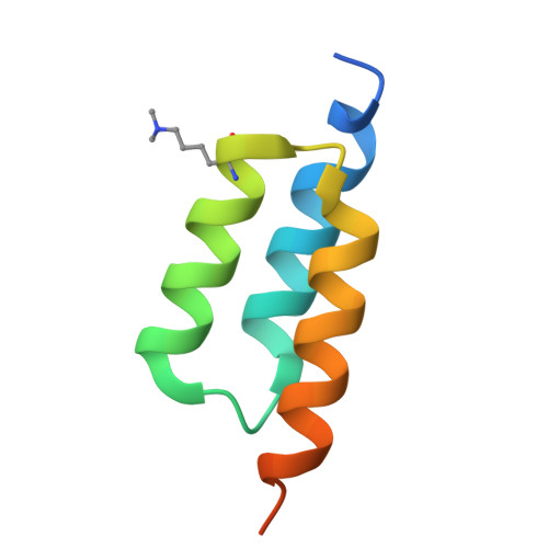
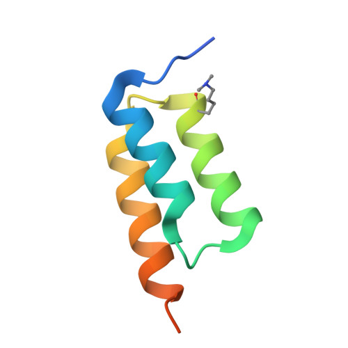
Coevolved affibody pair A6B6


Experimental Data Snapshot

  • Method: X-RAY DIFFRACTION
  • Resolution: 1.43 Å
  • R-Value Free: 
    0.214 (Depositor), 0.233 (DCC) 
  • R-Value Work: 
    0.187 (Depositor) 
  • R-Value Observed: 
    0.189 (Depositor) 

Starting Models: experimental
View more details

wwPDB Validation 3D Report Full Report

Validation slider image for 9NKQ

This is version 1.1 of the entry. See complete history

Literature

Structural ontogeny of protein-protein interactions.

Yang, A.Jiang, H.Jude, K.M.Akpinaroglu, D.Allenspach, S.Li, A.J.Bowden, J.Perez, C.P.Liu, L.Huang, P.S.Kortemme, T.Listgarten, J.Garcia, K.C.

(2026) Science 391: eadx6931-eadx6931

  • DOI: https://doi.org/10.1126/science.adx6931
  • Primary Citation Related Structures: 
    9NKM, 9NKN, 9NKO, 9NKP, 9NKQ, 9NKR, 9NKS

  • PubMed Abstract: 

    Understanding how protein binding sites evolve interactions with other proteins could hold clues to targeting "undruggable" surfaces. We used synthetic coevolution to engineer new interactions between naïve surfaces, simulating the de novo formation of protein complexes. We isolated seven distinct structural families of protein Z-domain complexes and found that synthetic complexes explore multiple shallow energy wells through ratchet-like docking modes, whereas complexes formed by natural binding sites converged in a deep energy well with a relatively fixed geometry. Epistasis analysis of a machine learning-estimated fitness landscape revealed "seed" contacts between binding partners that anchored the earliest stages of encounter complex formation. Our results suggest that "silent" surfaces have a shallower energy landscape than natural binding sites, disfavoring tight binding, likely owing to evolutionary counterselection.


  • Organizational Affiliation
    • Department of Molecular and Cellular Physiology, Stanford University School of Medicine, Stanford, CA, USA.

Macromolecule Content 

  • Total Structure Weight: 23.61 kDa 
  • Atom Count: 1,459 
  • Modeled Residue Count: 165 
  • Deposited Residue Count: 204 
  • Unique protein chains: 2

Macromolecules

Find similar proteins by:|  3D Structure
Entity ID: 1
MoleculeChains  Sequence LengthOrganismDetailsImage
A6 affibody68synthetic constructMutation(s): 0 
Find similar proteins by:|  3D Structure
Entity ID: 2
MoleculeChains  Sequence LengthOrganismDetailsImage
B6 affibodyB,
C [auth D]
68synthetic constructMutation(s): 0 

Small Molecules

Ligands 1 Unique
IDChains Name / Formula / InChI Key2D Diagram3D Interactions
SO4

Query on SO4


Download:Ideal Coordinates CCD File
D [auth A]SULFATE ION
O4 S
QAOWNCQODCNURD-UHFFFAOYSA-L
Modified Residues  1 Unique
IDChains TypeFormula2D DiagramParent
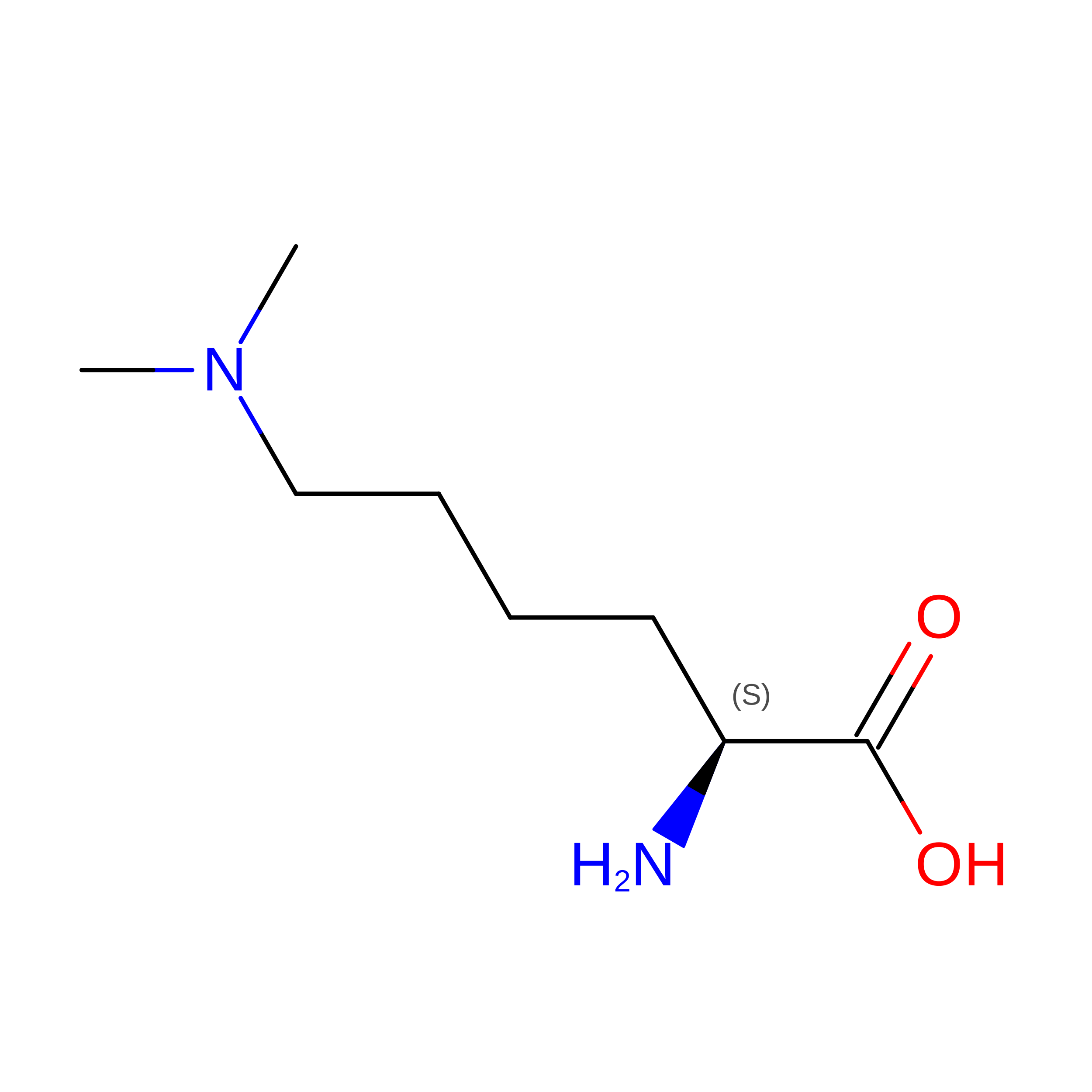
MLY
Query on MLY
A
L-PEPTIDE LINKINGC8 H18 N2 O2LYS

Experimental Data & Validation

Experimental Data

  • Method: X-RAY DIFFRACTION
  • Resolution: 1.43 Å
  • R-Value Free:  0.214 (Depositor), 0.233 (DCC) 
  • R-Value Work:  0.187 (Depositor) 
  • R-Value Observed: 0.189 (Depositor) 
Space Group: P 21 21 21
Unit Cell:
Length ( Å )Angle ( ˚ )
a = 44.393α = 90
b = 44.653β = 90
c = 92.514γ = 90
Software Package:
Software NamePurpose
PHENIXrefinement
XDSdata scaling
PHASERphasing
XDSdata reduction

Structure Validation

View Full Validation Report


Entry History 

& Funding Information

Deposition Data


Funding OrganizationLocationGrant Number
National Institutes of Health/National Institute of General Medical Sciences (NIH/NIGMS)United StatesGM150125
Howard Hughes Medical Institute (HHMI)United States--

Revision History  (Full details and data files)

  • Version 1.0: 2026-02-04
    Type: Initial release
  • Version 1.1: 2026-02-25
    Changes: Database references