9NIH | pdb_00009nih

Crystal structure of HLA-DR4 presenting citrullinated Tenascin C peptide


Experimental Data Snapshot

  • Method: X-RAY DIFFRACTION
  • Resolution: 2.40 Å
  • R-Value Free: 
    0.219 (Depositor), 0.218 (DCC) 
  • R-Value Work: 
    0.184 (Depositor), 0.184 (DCC) 
  • R-Value Observed: 
    0.186 (Depositor) 

Starting Model: experimental
View more details

wwPDB Validation 3D Report Full Report

Validation slider image for 9NIH

Ligand Structure Quality Assessment 


This is version 1.1 of the entry. See complete history

Literature

The molecular basis of T cell receptor recognition of citrullinated tenascin-C presented by HLA-DR4.

Dao, H.T.Loh, T.J.Sharma, R.K.Klareskog, L.Malmstrom, V.Reid, H.H.Rossjohn, J.Lim, J.J.

(2025) J Biological Chem 301: 110326-110326

  • DOI: https://doi.org/10.1016/j.jbc.2025.110326
  • Primary Citation Related Structures: 
    9NIG, 9NIH, 9NII

  • PubMed Abstract: 

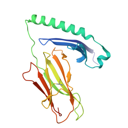
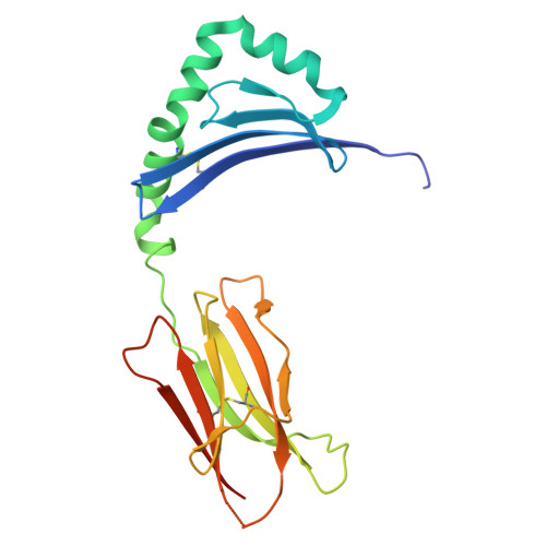
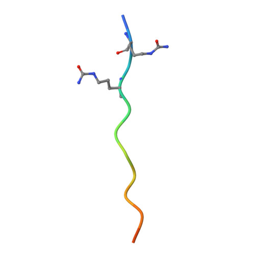
    CD4 + T cell autoreactivity against citrullinated (cit) self-epitopes presented by HLA-DRB1 is associated with rheumatoid arthritis (RA) pathogenesis. We understand the molecular bases of T cell receptor (TCR) recognition of cit-fibrinogen, cit-vimentin, and cit-α-enolase epitopes, and the role of citrulline in shaping TCR repertoire usage. Nevertheless, how TCRs recognize other cit-epitopes, including tenascin-C (TNC) and how alternative citrullination positions may modulate the T cell recognition remains unclear. Here, we examined TNC 1014,1016cit peptide, which contains citrullination at position P-1 and P2, to study the underlying TCR-HLA-DRB1 04:01-TNC 1014,1016cit molecular interactions. Crystal structure of HLA-DRB1 04:01 TNC1014,1016cit at 2.4 Å resolution revealed a conserved peptide binding register to the established HLA-DRB1 04:01-peptide structures, where both citrullines protruded upward. Next, we determined the crystal structure of a RA patient-derived TRAV35 + /TRBV10-2 + (PB) TCR in complex with HLA-DRB1 04:01 TNC1014,1016cit at 3.2 Å resolution. The CDR3α loop ( 109 VGNTN 113 ) of PB TCR formed a secondary helical conformation at the N-terminus of the peptide binding cleft, allowing extensive interactions between the P-1 and P2 citrullines of TNC 1014,1016cit peptide. Surface plasmon resonance, tetramer staining, and CD69 activation assays revealed that the PB TCR did not cross-react to other RA autoantigens, and the P-1-Cit, P2-Cit, and P5-Tyr of TNC 1014,1016cit are the key determinants underlying the strict specificity of the PB TCR. Collectively, we provide molecular insight into citrullination in modulating TCR recognition.


  • Organizational Affiliation
    • Infection and Immunity Program and Department of Biochemistry and Molecular Biology, Biomedicine Discovery Institute, Monash University, Clayton, Australia.

Macromolecule Content 

  • Total Structure Weight: 47.43 kDa 
  • Atom Count: 3,232 
  • Modeled Residue Count: 381 
  • Deposited Residue Count: 401 
  • Unique protein chains: 3

Macromolecules

Find similar proteins by:|  3D Structure
Entity ID: 1
MoleculeChains  Sequence LengthOrganismDetailsImage
HLA class II histocompatibility antigen, DR alpha chain189Homo sapiensMutation(s): 0 
Gene Names: HLA-DRAHLA-DRA1
UniProt & NIH Common Fund Data Resources
Find proteins for P01903 (Homo sapiens)
Explore P01903 
Go to UniProtKB:  P01903
PHAROS:  P01903
GTEx:  ENSG00000204287 
Entity Groups
Sequence Clusters30% Identity50% Identity70% Identity90% Identity95% Identity100% Identity
UniProt GroupP01903
Glycosylation
Glycosylation Sites: 1Go to GlyGen: P01903-1
Sequence Annotations
Expand
Reference Sequence
Find similar proteins by:|  3D Structure
Entity ID: 2
MoleculeChains  Sequence LengthOrganismDetailsImage
HLA class II histocompatibility antigen DR beta chain198Homo sapiensMutation(s): 0 
Gene Names: HLA-DRB1
UniProt
Find proteins for A0A1V1IGJ9 (Homo sapiens)
Explore A0A1V1IGJ9 
Go to UniProtKB:  A0A1V1IGJ9
Entity Groups
Sequence Clusters30% Identity50% Identity70% Identity90% Identity95% Identity100% Identity
UniProt GroupA0A1V1IGJ9
Glycosylation
Glycosylation Sites: 1
Sequence Annotations
Expand
Reference Sequence
Find similar proteins by:  Sequence   |   3D Structure  
Entity ID: 3
MoleculeChains  Sequence LengthOrganismDetailsImage
Tenascin14Homo sapiensMutation(s): 0 
UniProt & NIH Common Fund Data Resources
Find proteins for P24821 (Homo sapiens)
Explore P24821 
Go to UniProtKB:  P24821
PHAROS:  P24821
GTEx:  ENSG00000041982 
Entity Groups
Sequence Clusters30% Identity50% Identity70% Identity90% Identity95% Identity100% Identity
UniProt GroupP24821
Sequence Annotations
Expand
Reference Sequence

Experimental Data & Validation

Experimental Data

  • Method: X-RAY DIFFRACTION
  • Resolution: 2.40 Å
  • R-Value Free:  0.219 (Depositor), 0.218 (DCC) 
  • R-Value Work:  0.184 (Depositor), 0.184 (DCC) 
  • R-Value Observed: 0.186 (Depositor) 
Space Group: H 3
Unit Cell:
Length ( Å )Angle ( ˚ )
a = 119.3α = 90
b = 119.3β = 90
c = 73.273γ = 120
Software Package:
Software NamePurpose
PHENIXrefinement
XDSdata reduction
Aimlessdata scaling
PHASERphasing

Structure Validation

View Full Validation Report


Ligand Structure Quality Assessment 


Entry History 

& Funding Information

Deposition Data


Funding OrganizationLocationGrant Number
Not funded--

Revision History  (Full details and data files)

  • Version 1.0: 2025-05-21
    Type: Initial release
  • Version 1.1: 2025-12-03
    Changes: Database references