9N4U | pdb_00009n4u

Crystal structure of PAK1 bound to compound R1


Experimental Data Snapshot

  • Method: X-RAY DIFFRACTION
  • Resolution: 1.77 Å
  • R-Value Free: 
    0.203 (Depositor), 0.203 (DCC) 
  • R-Value Work: 
    0.173 (Depositor), 0.174 (DCC) 
  • R-Value Observed: 
    0.175 (Depositor) 

Starting Model: experimental
View more details

wwPDB Validation 3D Report Full Report

Validation slider image for 9N4U

Ligand Structure Quality Assessment 


This is version 1.1 of the entry. See complete history

Literature

Integrating Hydrogen Exchange with Molecular Dynamics for Improved Ligand Binding Predictions.

Walters, B.T.Patapoff, A.W.Kiefer, J.R.Wu, P.Wang, W.

(2025) J Chem Inf Model 65: 6144-6154

  • DOI: https://doi.org/10.1021/acs.jcim.5c00397
  • Primary Citation Related Structures: 
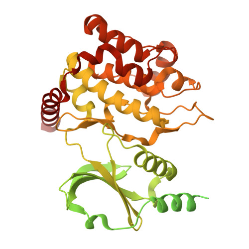
    9N48, 9N4U, 9N7R, 9N9X, 9NAC, 9NBS, 9NBX, 9NC2

  • PubMed Abstract: 

    We introduce hydrogen-exchange experimental structure prediction (HX-ESP), a method that integrates hydrogen exchange (HX) data with molecular dynamics (MD) simulations to accurately predict ligand binding modes, even for targets requiring significant conformational changes. Benchmarking HX-ESP by fitting two ligands to PAK1 and four ligands to MAP4K1 (HPK1) and comparing the results to X-ray crystallography structures, demonstrates that HX-ESP can identify binding modes across a range of affinities significantly outperforming flexible docking for ligands necessitating large conformational adjustments. By objectively guiding simulations with experimental HX data, HX-ESP overcomes the long time scales required for binding predictions using traditional MD. This advancement enhances the accuracy of computational modeling in drug discovery and thus will accelerate the development of effective therapeutics.


  • Organizational Affiliation
    • Department of Biochemistry and Cellular Pharmacology, Genentech, Inc., 1 DNA Way, South San Francisco, California 94080, United States.

Macromolecule Content 

  • Total Structure Weight: 123.59 kDa 
  • Atom Count: 5,092 
  • Modeled Residue Count: 579 
  • Deposited Residue Count: 1,074 
  • Unique protein chains: 1

Macromolecules

Find similar proteins by:|  3D Structure
Entity ID: 1
MoleculeChains  Sequence LengthOrganismDetailsImage
Glutathione S-transferase class-mu 26 kDa isozyme,Serine/threonine-protein kinase PAK 1
A, B
537Homo sapiensMutation(s): 0 
EC: 2.5.1.18 (PDB Primary Data), 2.7.11.1 (PDB Primary Data)
UniProt & NIH Common Fund Data Resources
Find proteins for P08515 (Schistosoma japonicum)
Explore P08515 
Go to UniProtKB:  P08515
Find proteins for Q13153 (Homo sapiens)
Explore Q13153 
Go to UniProtKB:  Q13153
PHAROS:  Q13153
GTEx:  ENSG00000149269 
Entity Groups
Sequence Clusters30% Identity50% Identity70% Identity90% Identity95% Identity100% Identity
UniProt GroupsQ13153P08515
Sequence Annotations
Expand
Reference Sequence

Small Molecules

Ligands 1 Unique
IDChains Name / Formula / InChI Key2D Diagram3D Interactions
A1BV8
(Subject of Investigation/LOI)

Query on A1BV8


Download:Ideal Coordinates CCD File
C [auth A],
D [auth B]
(3M)-3-[(4P)-2-chloro-4-(6-methylpyridin-2-yl)phenyl]-1-{2-[2-(dimethylamino)ethoxy]ethyl}-1,6-naphthyridin-2(1H)-one
C26 H27 Cl N4 O2
NPOXYSJSLYNRSE-UHFFFAOYSA-N

Experimental Data & Validation

Experimental Data

  • Method: X-RAY DIFFRACTION
  • Resolution: 1.77 Å
  • R-Value Free:  0.203 (Depositor), 0.203 (DCC) 
  • R-Value Work:  0.173 (Depositor), 0.174 (DCC) 
  • R-Value Observed: 0.175 (Depositor) 
Space Group: P 1 21 1
Unit Cell:
Length ( Å )Angle ( ˚ )
a = 62.386α = 90
b = 80.728β = 106.92
c = 65.893γ = 90
Software Package:
Software NamePurpose
PHENIXrefinement
HKL-2000data scaling
HKL-2000data reduction
PHASERphasing

Structure Validation

View Full Validation Report


Ligand Structure Quality Assessment 


Entry History 

& Funding Information

Deposition Data


Funding OrganizationLocationGrant Number
Other privateUnited States--

Revision History  (Full details and data files)

  • Version 1.0: 2025-06-25
    Type: Initial release
  • Version 1.1: 2025-07-02
    Changes: Database references