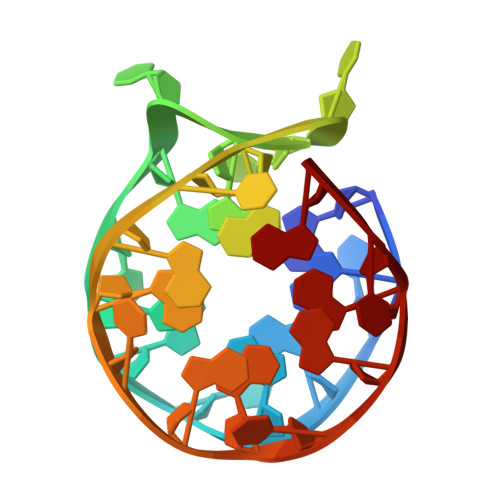
Preservation of left-handed fold and interaction with N-methylmesoporphyrin IX by two-block sequences with left-hand folding potential
Hendrickson, A.D., Xing, E.R., Yatsunyk, L.A.To be published.
Experimental Data Snapshot
Starting Model: experimental
View more details
| Ligands 5 Unique | |||||
|---|---|---|---|---|---|
| ID | Chains | Name / Formula / InChI Key | 2D Diagram | 3D Interactions | |
| SPM Download:Ideal Coordinates CCD File | K [auth B], L [auth B] | SPERMINE C10 H26 N4 PFNFFQXMRSDOHW-UHFFFAOYSA-N |  | ||
| PG4 Download:Ideal Coordinates CCD File | Q [auth C], R [auth C] | TETRAETHYLENE GLYCOL C8 H18 O5 UWHCKJMYHZGTIT-UHFFFAOYSA-N |  | ||
| PGE Download:Ideal Coordinates CCD File | S [auth C], W [auth D] | TRIETHYLENE GLYCOL C6 H14 O4 ZIBGPFATKBEMQZ-UHFFFAOYSA-N |  | ||
| PEG Download:Ideal Coordinates CCD File | M [auth B] | DI(HYDROXYETHYL)ETHER C4 H10 O3 MTHSVFCYNBDYFN-UHFFFAOYSA-N |  | ||
| K Download:Ideal Coordinates CCD File | E [auth A] F [auth A] G [auth A] H [auth B] I [auth B] | POTASSIUM ION K NPYPAHLBTDXSSS-UHFFFAOYSA-N |  | ||
| Length ( Å ) | Angle ( ˚ ) |
|---|---|
| a = 104.649 | α = 90 |
| b = 104.649 | β = 90 |
| c = 65.365 | γ = 120 |
| Software Name | Purpose |
|---|---|
| PHENIX | refinement |
| autoPROC | data reduction |
| autoPROC | data scaling |
| PHENIX | phasing |
| Funding Organization | Location | Grant Number |
|---|---|---|
| National Institutes of Health/National Cancer Institute (NIH/NCI) | United States | 1R15CA253134 |