Download Mendeley
Structure and inhibition of diaminopimelic acid epimerase by slow-binding alpha-methyl amino acids.
Lamer, T., Chen, P., Catenza, K., Perov, I., L, B., Hsiao, Y.T., Van Oers, T.J., Lemieux, M.J., Vederas, J.C.(2025) Protein Sci 34: e70139-e70139
- PubMed: 40299312 Search on PubMedSearch on PubMed Central
- DOI: https://doi.org/10.1002/pro.70139
- Primary Citation Related Structures:
9MRO, 9MRP, 9MRV - PubMed Abstract:
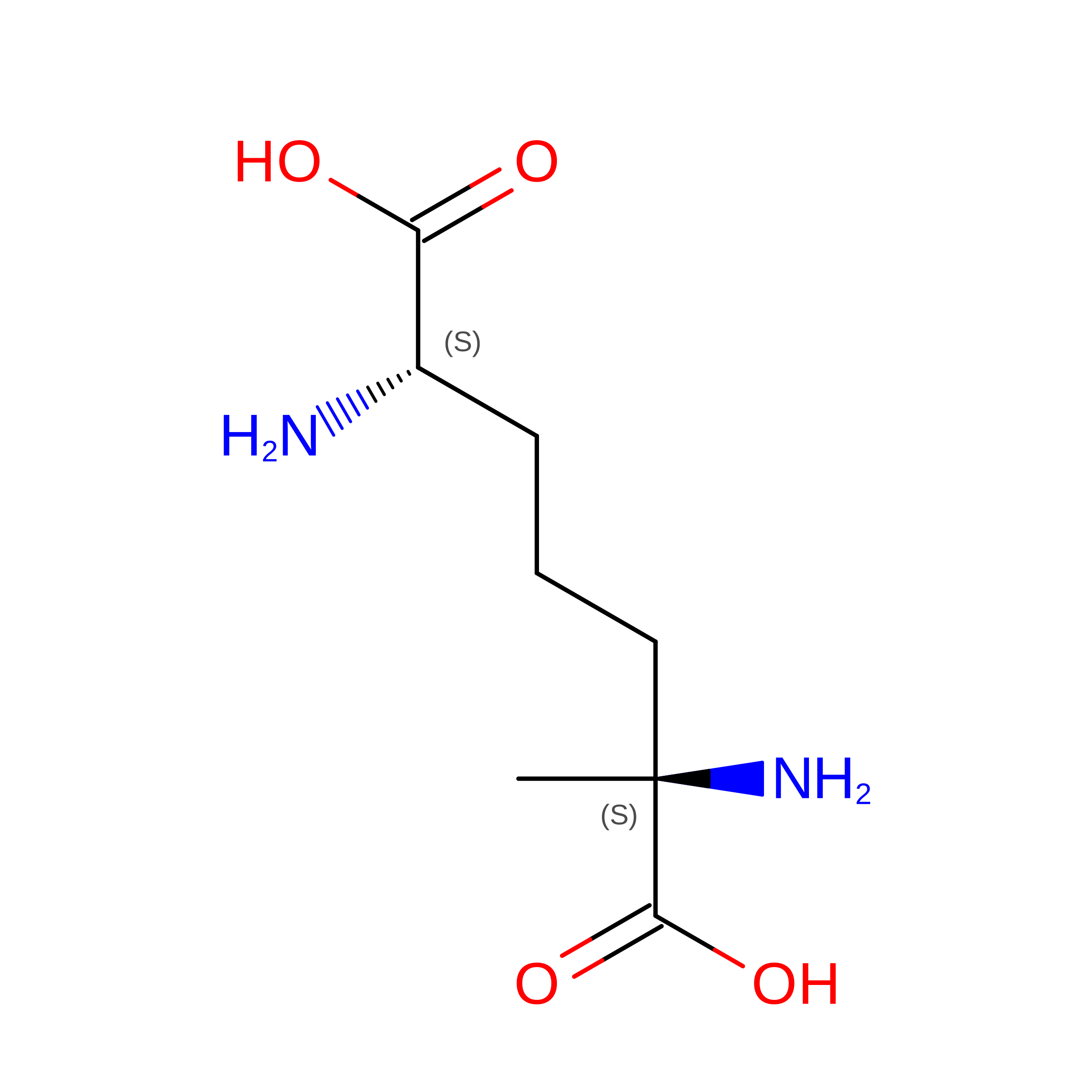
Cofactor-independent racemases and epimerases produce D-amino acids from their L-isomers for a variety of biological processes. These enzymes operate via an unusual mechanism that relies on an active site cysteine thiolate (pK a ~ 8.5) to deprotonate an amino acid α-carbon (pK a ~ 29) and are of interest not only because of their biocatalytic potential for D-amino acid production, but also because many play key roles in biology and are antibiotic targets. However, obtaining crystal structures of these enzymes, especially in their closed, substrate- or inhibitor-bound conformations, is difficult. In this work, we characterized diaminopimelic acid (DAP) epimerase from the cyanobacterium Anabaena. DAP epimerase has long been of interest as an antibiotic target as it converts L,L-DAP to D,L-DAP for lysine and peptidoglycan biosynthesis. We solved three crystal structures of this enzyme in its closed, inhibitor-bound conformation, up to a resolution of 1.5 Å. Two structures show the enzyme covalently bound through its catalytic cysteine residues to previously reported aziridine-based inhibitors. One structure unexpectedly shows the enzyme bound to a different compound, D,L-α-methylDAP, presumably produced as a synthetic byproduct. Stereoselective synthesis of L,L- and D,L-α-methylDAP followed by inhibition assays shows that these compounds are slow-binding inhibitors of DAP epimerase. α-MethylDAP inhibitors provide a more accessible alternative to aziridine-based inhibitors to obtain crystal structures of DAP epimerase in its closed conformation. Comparisons of bacterial, cyanobacterial, and plant DAP epimerases provided here offer new insights into functional and structural differences between these enzymes.
- Department of Chemistry, University of Alberta, Edmonton, Alberta, Canada.
Organizational Affiliation: