9MOH | pdb_00009moh

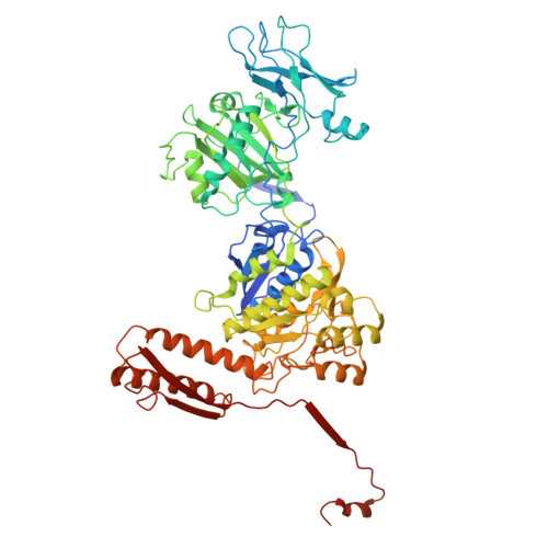
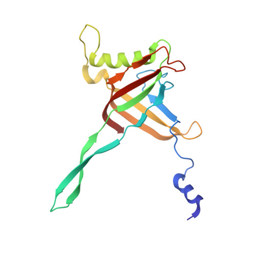
Structure of the middle part of the bacteriophage T4 tail


Experimental Data Snapshot

  • Method: ELECTRON MICROSCOPY
  • Resolution: 3.40 Å
  • Aggregation State: PARTICLE 
  • Reconstruction Method: SINGLE PARTICLE 

wwPDB Validation 3D Report Full Report

Validation slider image for 9MOH

This is version 1.1 of the entry. See complete history

Literature

In situ structures of the portal-neck-tail complex of bacteriophage T4 inform a viral genome positioning mechanism.

Fokine, A.Zhu, J.Klose, T.Vago, F.Arnaud, C.A.Wang, Z.Khare, B.Rossmann, M.G.Chen, Z.Sun, L.Fang, Q.Kuhn, R.J.Rao, V.B.

(2026) Nat Commun 17

  • DOI: https://doi.org/10.1038/s41467-026-69106-8
  • Primary Citation Related Structures: 
    9MKB, 9MOF, 9MOG, 9MOH

  • PubMed Abstract: 

    The post-genome packaging mechanisms that govern the assembly of an infectious virion are poorly understood in bacteriophages and other viruses. Here, our near-atomic resolution cryo-EM structural analyses uncovered an assembly- and conformation-driven genome positioning mechanism in the tailed bacteriophage T4. We show that following headful packaging, which generates a pressurized head, a global conformational change occurs in the portal structure, probably triggering packaging termination and ejection of the packaging motor. Our high-resolution structures of the neck of the virion further show that the neck undergoes conformational changes upon docking of a pre-assembled tail onto the sealed neck, which then opens a genome-gate. Driven by the pressure of the packaged DNA, the genome travels through open neck channels, binds and compresses the resident tape-measure protein, and halts at the bottom of the second topmost disk of the tail tube. Pressure-suspended within the virion's innermost tunnel and secured by a baseplate plug, the genome remains poised to flow through the tunnel into a host cell upon receiving the host receptor recognition signal.


  • Organizational Affiliation
    • Department of Biological Sciences, Purdue University, West Lafayette, IN, USA. afokine@purdue.edu.

Macromolecule Content 

  • Total Structure Weight: 7,434.69 kDa 
  • Atom Count: 521,712 
  • Modeled Residue Count: 67,734 
  • Deposited Residue Count: 68,070 
  • Unique protein chains: 2

Macromolecules

Find similar proteins by:|  3D Structure
Entity ID: 1
MoleculeChains  Sequence LengthOrganismDetailsImage
gp18, tail sheath protein659Escherichia phage T4Mutation(s): 0 
UniProt
Find proteins for A0A7S9SVW9 (Escherichia phage T4)
Explore A0A7S9SVW9 
Go to UniProtKB:  A0A7S9SVW9
Entity Groups
Sequence Clusters30% Identity50% Identity70% Identity90% Identity95% Identity100% Identity
UniProt GroupA0A7S9SVW9
Sequence Annotations
Expand
Reference Sequence
Find similar proteins by:|  3D Structure
Entity ID: 2
MoleculeChains  Sequence LengthOrganismDetailsImage
gp19, tail tube protein163Escherichia phage T4Mutation(s): 0 
UniProt
Find proteins for A0A7S9SVI2 (Escherichia phage T4)
Explore A0A7S9SVI2 
Go to UniProtKB:  A0A7S9SVI2
Entity Groups
Sequence Clusters30% Identity50% Identity70% Identity90% Identity95% Identity100% Identity
UniProt GroupA0A7S9SVI2
Sequence Annotations
Expand
Reference Sequence

Experimental Data & Validation

Experimental Data

  • Method: ELECTRON MICROSCOPY
  • Resolution: 3.40 Å
  • Aggregation State: PARTICLE 
  • Reconstruction Method: SINGLE PARTICLE 
EM Software:
TaskSoftware PackageVersion
MODEL REFINEMENTPHENIX1.21.1_5286

Structure Validation

View Full Validation Report


Entry History 

& Funding Information

Deposition Data


Funding OrganizationLocationGrant Number
National Institutes of Health/National Institute Of Allergy and Infectious Diseases (NIH/NIAID)United StatesAI081726

Revision History  (Full details and data files)

  • Version 1.0: 2026-01-28
    Type: Initial release
  • Version 1.1: 2026-08-12
    Changes: Data collection, Database references