9MO7 | pdb_00009mo7

Structure of native murine cardiac thin filament at pCa=5.8 in Ca2+-bound fully activated state (lower strand)

  • Classification: MOTOR PROTEIN
  • Organism(s): Mus musculus
  • Mutation(s): No 

  • Deposited: 2024-12-25 Released: 2025-05-28 
  • Deposition Author(s): Risi, C.M., Galkin, V.E.
  • Funding Organization(s): National Institutes of Health/National Heart, Lung, and Blood Institute (NIH/NHLBI)

Experimental Data Snapshot

  • Method: ELECTRON MICROSCOPY
  • Resolution: 5.20 Å
  • Aggregation State: FILAMENT 
  • Reconstruction Method: SINGLE PARTICLE 

Starting Models: experimental, in silico
View more details

wwPDB Validation 3D Report Full Report

Validation slider image for 9MO7

This is version 1.1 of the entry. See complete history

Literature

The role of the troponin T interactions with actin in regulation of cardiac thin filament revealed by the troponin T pathogenic variant Ile79Asn.

Risi, C.M.Landim-Vieira, M.Belknap, B.Chase, P.B.Pinto, J.R.Galkin, V.E.

(2025) J Mol Cell Cardiol 204: 55-67

  • DOI: https://doi.org/10.1016/j.yjmcc.2025.05.005
  • Primary Citation Related Structures: 
    9E2E, 9MO4, 9MO5, 9MO6, 9MO7, 9MO8, 9MO9, 9MOA, 9MOB, 9MOC, 9MOD, 9MOI, 9MOK, 9MOL, 9MOM, 9MON, 9MOO, 9MOP, 9MOU, 9MOW, 9MOX

  • PubMed Abstract: 

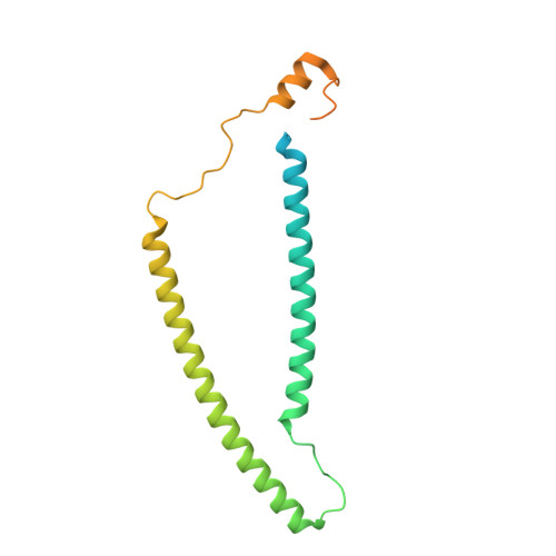
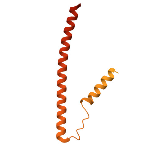
    Cardiac muscle contraction/relaxation cycle depends on the rising and falling Ca 2+ levels in sarcomeres that control the extent of interactions between myosin-based thick and actin-based thin filaments. Cardiac thin filament (cTF) consists of actin, tropomyosin (Tm) that regulates myosin binding to actin, and troponin complex that governs Tm position upon Ca 2+ -binding. Troponin has three subunits - Ca 2+ -binding troponin C (TnC), Tm stabilizing troponin T (TnT), and inhibitory troponin I (TnI). TnT N-terminus (TnT1) interactions with actin stabilize the inhibited state of cTF. TnC, TnI, and Tm work in concert to control actomyosin interactions. Cryo-electron microscopy (cryo-EM) provided factual structures of healthy cTF, but structures of cTF carrying missense mutations linked to human cardiomyopathy are unknown. Variant Ile79Asn in human cardiac TnT (TnT-I79N) increases myofilament Ca 2+ sensitivity and slows cross-bridge kinetics, leading to severe hypertrophic/restrictive cardiomyopathy. Here, we used TnT-I79N mutation as a tool to examine the role of TnT1 in the complex mechanism of cTF regulation. Comparison of the cryo-EM structures of murine wild type and TnT-I79N native cTFs at systolic Ca 2+ levels (pCa = 5.8) demonstrates that TnT-I79N causes 1) dissociation of the TnT1 loop from its actin interface that results in Tm release to a more activated position, 2) reduced interaction of TnI C-terminus with actin-Tm, and 3) increased frequency of Ca 2+ -bound regulatory units. Our data indicate that the TnT1 loop is a crucial element of the allosteric regulatory network that couples Tn subunits and Tm to maintain adequate cTF response to physiological Ca 2+ levels during a heartbeat.


  • Organizational Affiliation
    • Department of Biomedical and Translational Sciences, Macon & Joan Brock Virginia Health Sciences at Old Dominion University, Norfolk, VA 23507, USA.

Macromolecule Content 

  • Total Structure Weight: 498.14 kDa 
  • Atom Count: 25,730 
  • Modeled Residue Count: 3,228 
  • Deposited Residue Count: 4,352 
  • Unique protein chains: 5

Macromolecules

Find similar proteins by:|  3D Structure
Entity ID: 1
MoleculeChains  Sequence LengthOrganismDetailsImage
Actin, alpha cardiac muscle 1
A, B, C, D, E
A, B, C, D, E, F
377Mus musculusMutation(s): 0 
EC: 3.6.4
UniProt & NIH Common Fund Data Resources
Find proteins for P68033 (Mus musculus)
Explore P68033 
Go to UniProtKB:  P68033
IMPC:  MGI:87905
Entity Groups
Sequence Clusters30% Identity50% Identity70% Identity90% Identity95% Identity100% Identity
UniProt GroupP68033
Sequence Annotations
Expand
Reference Sequence
Find similar proteins by:|  3D Structure
Entity ID: 2
MoleculeChains  Sequence LengthOrganismDetailsImage
Troponin C, slow skeletal and cardiac muscles161Mus musculusMutation(s): 0 
UniProt & NIH Common Fund Data Resources
Find proteins for P19123 (Mus musculus)
Explore P19123 
Go to UniProtKB:  P19123
IMPC:  MGI:98779
Entity Groups
Sequence Clusters30% Identity50% Identity70% Identity90% Identity95% Identity100% Identity
UniProt GroupP19123
Sequence Annotations
Expand
Reference Sequence
Find similar proteins by:|  3D Structure
Entity ID: 3
MoleculeChains  Sequence LengthOrganismDetailsImage
Troponin I, cardiac muscle211Mus musculusMutation(s): 0 
UniProt
Find proteins for P48787 (Mus musculus)
Explore P48787 
Go to UniProtKB:  P48787
Entity Groups
Sequence Clusters30% Identity50% Identity70% Identity90% Identity95% Identity100% Identity
UniProt GroupP48787
Sequence Annotations
Expand
Reference Sequence
Find similar proteins by:|  3D Structure
Entity ID: 4
MoleculeChains  Sequence LengthOrganismDetailsImage
Isoform A2 of Troponin T, cardiac muscle
I, J
291Mus musculusMutation(s): 0 
UniProt
Find proteins for P50752 (Mus musculus)
Explore P50752 
Go to UniProtKB:  P50752
Entity Groups
Sequence Clusters30% Identity50% Identity70% Identity90% Identity95% Identity100% Identity
UniProt GroupP50752
Sequence Annotations
Expand
Reference Sequence
Find similar proteins by:|  3D Structure
Entity ID: 5
MoleculeChains  Sequence LengthOrganismDetailsImage
Tropomyosin alpha-1 chain
K, L, M, N
284Mus musculusMutation(s): 0 
UniProt & NIH Common Fund Data Resources
Find proteins for P58771 (Mus musculus)
Explore P58771 
Go to UniProtKB:  P58771
IMPC:  MGI:98809
Entity Groups
Sequence Clusters30% Identity50% Identity70% Identity90% Identity95% Identity100% Identity
UniProt GroupP58771
Sequence Annotations
Expand
Reference Sequence

Small Molecules

Ligands 3 Unique
IDChains Name / Formula / InChI Key2D Diagram3D Interactions
ADP

Query on ADP


Download:Ideal Coordinates CCD File
O [auth A]
Q [auth B]
S [auth C]
U [auth D]
W [auth E]
O [auth A],
Q [auth B],
S [auth C],
U [auth D],
W [auth E],
Y [auth F]
ADENOSINE-5'-DIPHOSPHATE
C10 H15 N5 O10 P2
XTWYTFMLZFPYCI-KQYNXXCUSA-N
CA

Query on CA


Download:Ideal Coordinates CCD File
AA [auth G],
BA [auth G],
CA [auth G]
CALCIUM ION
Ca
BHPQYMZQTOCNFJ-UHFFFAOYSA-N
MG

Query on MG


Download:Ideal Coordinates CCD File
P [auth A]
R [auth B]
T [auth C]
V [auth D]
X [auth E]
P [auth A],
R [auth B],
T [auth C],
V [auth D],
X [auth E],
Z [auth F]
MAGNESIUM ION
Mg
JLVVSXFLKOJNIY-UHFFFAOYSA-N

Experimental Data & Validation

Experimental Data

  • Method: ELECTRON MICROSCOPY
  • Resolution: 5.20 Å
  • Aggregation State: FILAMENT 
  • Reconstruction Method: SINGLE PARTICLE 
EM Software:
TaskSoftware PackageVersion
MODEL REFINEMENTPHENIX
RECONSTRUCTIONRELION

Structure Validation

View Full Validation Report


Entry History 

& Funding Information

Deposition Data


Funding OrganizationLocationGrant Number
National Institutes of Health/National Heart, Lung, and Blood Institute (NIH/NHLBI)United StatesR01 HL160966

Revision History  (Full details and data files)

  • Version 1.0: 2025-05-28
    Type: Initial release
  • Version 1.1: 2025-07-02
    Changes: Data collection, Database references