9LNK | pdb_00009lnk

Crystal structure of VANC21 in complex with its target DNA (native).


Experimental Data Snapshot

  • Method: X-RAY DIFFRACTION
  • Resolution: 2.55 Å
  • R-Value Free: 
    0.255 (Depositor), 0.255 (DCC) 
  • R-Value Work: 
    0.221 (Depositor), 0.221 (DCC) 
  • R-Value Observed: 
    0.223 (Depositor) 

Starting Model: experimental
View more details

wwPDB Validation 3D Report Full Report

Validation slider image for 9LNK

This is version 1.1 of the entry. See complete history

Literature

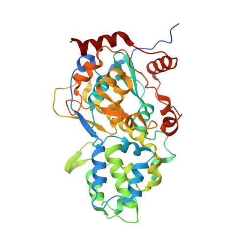
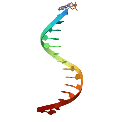
Structure and evolution of the sequence-specific anti-silencing factor VANC21 and its target DNA.

Tanaka, Y.Osakabe, A.Shihoya, W.Hirano, H.Itoh, Y.Kakutani, T.Nureki, O.

(2026) Genes Genet Syst 101

  • DOI: https://doi.org/10.1266/ggs.25-00096
  • Primary Citation Related Structures: 
    9LNJ, 9LNK

  • PubMed Abstract: 

    VANDAL family transposons are DNA transposons prevalent in Arabidopsis and related plants. A notable feature of VANDAL is that they can overcome epigenetic silencing from the host, using a VANC protein encoded in each VANDAL members: VANC21 protein encoded in VANDAL21 specifically accumulates on its target DNA motifs condensed in the non-coding regions of this TE and induces loss of DNA methylation, transcriptional derepression, and mobilization of them. In this study, to elucidate the mechanism of how VANC subtypes have diverged to bind specifically to their own target motifs in their cognate VANDAL subfamilies, we determined the crystal structure of VANC21 in complex with its target DNA at 2.0 Å resolution. The VANC structure adopts a globular novel fold with a Zn ion coordinated at the DNA-binding site. Interestingly, most DNA-interacting VANC residues are located in the loops but not in the conserved regions among VANC subtypes. This observation suggests that the high variability of DNA-interacting regions of VANC proteins brought about the co-evolution of VANCs and their target sequences. This rapid differentiation by co-evolution enabled VANDAL family TEs to proliferate while avoiding deleterious effects on the host fitness. Therefore, our findings help understand the adaptive evolutionary strategy for the survival of parasitic sequences.


  • Organizational Affiliation
    • Department of Biological Sciences, Graduate School of Sciences, The University of Tokyo.

Macromolecule Content 

  • Total Structure Weight: 53.46 kDa 
  • Atom Count: 3,619 
  • Modeled Residue Count: 392 
  • Deposited Residue Count: 413 
  • Unique protein chains: 1
  • Unique nucleic acid chains: 2

Macromolecules


Find similar proteins by:|  3D Structure
Entity ID: 1
MoleculeChains  Sequence LengthOrganismDetailsImage
VANC21383Arabidopsis thalianaMutation(s): 0 
UniProt
Find proteins for O80464 (Arabidopsis thaliana)
Explore O80464 
Go to UniProtKB:  O80464
Entity Groups
Sequence Clusters30% Identity50% Identity70% Identity90% Identity95% Identity100% Identity
UniProt GroupO80464
Sequence Annotations
Expand
Reference Sequence
Find similar nucleic acids by:  Sequence
Entity ID: 2
MoleculeChains LengthOrganismImage
VANC21 binding DNA fragment (motif strand)15Arabidopsis thaliana
Sequence Annotations
Expand
Reference Sequence
Find similar nucleic acids by:  Sequence
Entity ID: 3
MoleculeChains LengthOrganismImage
VANC21 binding DNA fragment (complementary strand)15Arabidopsis thaliana
Sequence Annotations
Expand
Reference Sequence

Small Molecules

Experimental Data & Validation

Experimental Data

  • Method: X-RAY DIFFRACTION
  • Resolution: 2.55 Å
  • R-Value Free:  0.255 (Depositor), 0.255 (DCC) 
  • R-Value Work:  0.221 (Depositor), 0.221 (DCC) 
  • R-Value Observed: 0.223 (Depositor) 
Space Group: P 21 21 21
Unit Cell:
Length ( Å )Angle ( ˚ )
a = 52.64α = 90
b = 96.85β = 90
c = 167.39γ = 90
Software Package:
Software NamePurpose
PHENIXrefinement
XDSdata reduction
XDSdata scaling
PHASERphasing

Structure Validation

View Full Validation Report


Entry History 

& Funding Information

Deposition Data


Funding OrganizationLocationGrant Number
Japan Society for the Promotion of Science (JSPS)Japan--

Revision History  (Full details and data files)

  • Version 1.0: 2025-12-10
    Type: Initial release
  • Version 1.1: 2026-02-18
    Changes: Database references