9LMT | pdb_00009lmt

Crystal structure of variant FAST-ACC-A248E


Experimental Data Snapshot

  • Method: X-RAY DIFFRACTION
  • Resolution: 1.50 Å
  • R-Value Free: 
    0.162 (Depositor), 0.162 (DCC) 
  • R-Value Work: 
    0.134 (Depositor), 0.134 (DCC) 
  • R-Value Observed: 
    0.135 (Depositor) 

Starting Model: experimental
View more details

wwPDB Validation 3D Report Full Report

Validation slider image for 9LMT

This is version 1.1 of the entry. See complete history

Literature

Combined approaches to enhance the Pichia pastoris-expressed PET hydrolase.

Li, X.Huang, J.W.Ning, Z.Huang, S.Zeng, C.Zeng, Z.Ji, R.Peng, R.Liu, X.Min, J.Chen, C.C.Guo, R.T.

(2025) Int J Biol Macromol 320: 145862-145862

  • DOI: https://doi.org/10.1016/j.ijbiomac.2025.145862
  • Primary Citation Related Structures: 
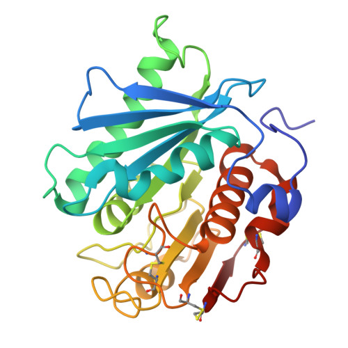
    9LMS, 9LMT, 9LMU, 9LMV, 9LMW, 9LMX

  • PubMed Abstract: 

    Enzymatic degradation of polyethylene terephthalate (PET) provides a sustainable and promising strategy for the recycling of plastic waste. Herein, we employed site-directed mutagenesis and machine learning methods to further enhance the performance of an efficient mutant of IsPETase, FAST-PETase-N212A, and resulting in seven variants with enhanced activity. We also found that the α3-β5 loop containing the T140D mutation plays a significant role in both type I and type II cutinases. Subsequently, we determined the complex structures of two activity-elevated mutants with the PET analogue mono(2-hydroxyethyl)terephthalic acid, revealing a different binding mode. Finally, to facilitate the industrial application of PET hydrolases, we exploited the industrial strain Pichia pastoris to express the activity-enhanced mutants. Compared with E. coli-produced proteins, these mutants expressed by P. pastoris exhibited higher activity and thermal stability.


  • Organizational Affiliation
    • Zhejiang Key Laboratory of Medical Epigenetics, Hubei Hongshan Laboratory, Department of Immunology and Pathogen Biology, School of Basic Medical Sciences, Hangzhou Normal University, 311121 Hangzhou, China; State Key Laboratory of Biocatalysis and Enzyme Engineering, School of Life Sciences, Hubei University, Wuhan 430062, China.

Macromolecule Content 

  • Total Structure Weight: 27.39 kDa 
  • Atom Count: 2,254 
  • Modeled Residue Count: 262 
  • Deposited Residue Count: 262 
  • Unique protein chains: 1

Macromolecules

Find similar proteins by:|  3D Structure
Entity ID: 1
MoleculeChains  Sequence LengthOrganismDetailsImage
Poly(ethylene terephthalate) hydrolase262Piscinibacter sakaiensisMutation(s): 1 
Gene Names: ISF6_4831
EC: 3.1.1.101
UniProt
Find proteins for A0A0K8P6T7 (Piscinibacter sakaiensis)
Explore A0A0K8P6T7 
Go to UniProtKB:  A0A0K8P6T7
Entity Groups
Sequence Clusters30% Identity50% Identity70% Identity90% Identity95% Identity100% Identity
UniProt GroupA0A0K8P6T7
Sequence Annotations
Expand
Reference Sequence

Experimental Data & Validation

Experimental Data

  • Method: X-RAY DIFFRACTION
  • Resolution: 1.50 Å
  • R-Value Free:  0.162 (Depositor), 0.162 (DCC) 
  • R-Value Work:  0.134 (Depositor), 0.134 (DCC) 
  • R-Value Observed: 0.135 (Depositor) 
Space Group: P 21 21 21
Unit Cell:
Length ( Å )Angle ( ˚ )
a = 51.026α = 90
b = 51.61β = 90
c = 84.496γ = 90
Software Package:
Software NamePurpose
REFMACrefinement
HKL-2000data scaling
HKL-2000data reduction
PHASERphasing

Structure Validation

View Full Validation Report


Entry History 

& Funding Information

Deposition Data


Funding OrganizationLocationGrant Number
National Natural Science Foundation of China (NSFC)China--

Revision History  (Full details and data files)

  • Version 1.0: 2025-07-23
    Type: Initial release
  • Version 1.1: 2025-07-30
    Changes: Database references