9LFU | pdb_00009lfu

Crystal structure of human RIP3 kinase domain complexed with LK01003


Experimental Data Snapshot

  • Method: X-RAY DIFFRACTION
  • Resolution: 2.93 Å
  • R-Value Free: 
    0.319 (Depositor), 0.307 (DCC) 
  • R-Value Work: 
    0.258 (Depositor) 
  • R-Value Observed: 
    0.261 (Depositor) 

Starting Model: experimental
View more details

wwPDB Validation 3D Report Full Report

Validation slider image for 9LFU

Ligand Structure Quality Assessment 


This is version 1.1 of the entry. See complete history

Literature

Structure-based design of potent and selective inhibitors targeting RIPK3 for eliminating on-target toxicity in vitro.

Su, H.Chen, G.Xie, H.Li, W.Xiong, M.He, J.Hu, H.Zhao, W.Shao, Q.Li, M.Zhao, Q.Xu, Y.

(2025) Nat Commun 16: 4288-4288

  • DOI: https://doi.org/10.1038/s41467-025-59432-8
  • Primary Citation Related Structures: 
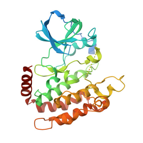
    9IWW, 9IWX, 9IWY, 9IWZ, 9IX0, 9IX1, 9IX2, 9IX3, 9LFU, 9LFV, 9LFW

  • PubMed Abstract: 

    The essential role of RIPK3 in necroptosis makes its inhibition a promising therapeutic strategy. However, the development of RIPK3 inhibitors has been hampered by on-target apoptosis and limited kinase selectivity. Inspired by the R69H mutation, which prevents on-target apoptosis by disrupting RIPK3 dimerization, we design LK-series inhibitors that effectively inhibit RIPK3 in biochemical assays and block TNF-α-induced necroptosis in both mouse L929 and human HT29 cells without inducing apoptosis. The representative compound, LK01003, shows high selectivity across a panel of 379 kinases. Our structural studies reveal that LK compounds act as Type I 1/2 inhibitors, engaging a unique hydrophobic site and stabilizing an inactive conformation of RIPK3. Moreover, several type II inhibitors are also revealed to maintain RIPK3 in the inactive conformation and do not induce on-target apoptosis. These findings suggest a promising strategy for rational design of safe and selective inhibitors by locking the inactive conformation of RIPK3.


  • Organizational Affiliation
    • State Key Laboratory of Drug Research, Shanghai Institute of Materia Medica, University of Chinese Academy of Sciences, Shanghai, 201203, China. suhaixia1@simm.ac.cn.

Macromolecule Content 

  • Total Structure Weight: 78.14 kDa 
  • Atom Count: 4,540 
  • Modeled Residue Count: 570 
  • Deposited Residue Count: 692 
  • Unique protein chains: 1

Macromolecules

Find similar proteins by:|  3D Structure
Entity ID: 1
MoleculeChains  Sequence LengthOrganismDetailsImage
Receptor-interacting serine/threonine-protein kinase 3
A, B
346Homo sapiensMutation(s): 2 
Gene Names: RIPK3RIP3
EC: 2.7.11.1
UniProt & NIH Common Fund Data Resources
Find proteins for Q9Y572 (Homo sapiens)
Explore Q9Y572 
Go to UniProtKB:  Q9Y572
PHAROS:  Q9Y572
GTEx:  ENSG00000129465 
Entity Groups
Sequence Clusters30% Identity50% Identity70% Identity90% Identity95% Identity100% Identity
UniProt GroupQ9Y572
Sequence Annotations
Expand
Reference Sequence

Small Molecules

Ligands 1 Unique
IDChains Name / Formula / InChI Key2D Diagram3D Interactions
A1D97

Query on A1D97


Download:Ideal Coordinates CCD File
C [auth A],
D [auth B]
2-cyclopentyl-~{N}-(6-propan-2-ylsulfonylquinolin-4-yl)-1,3-benzothiazol-5-amine
C24 H25 N3 O2 S2
PVKIFNZMYGMWPD-UHFFFAOYSA-N

Experimental Data & Validation

Experimental Data

  • Method: X-RAY DIFFRACTION
  • Resolution: 2.93 Å
  • R-Value Free:  0.319 (Depositor), 0.307 (DCC) 
  • R-Value Work:  0.258 (Depositor) 
  • R-Value Observed: 0.261 (Depositor) 
Space Group: P 64
Unit Cell:
Length ( Å )Angle ( ˚ )
a = 129.203α = 90
b = 129.203β = 90
c = 79.429γ = 120
Software Package:
Software NamePurpose
PHENIXrefinement
Aimlessdata scaling
XDSdata reduction
PHASERphasing

Structure Validation

View Full Validation Report


Ligand Structure Quality Assessment 


Entry History 

& Funding Information

Deposition Data


Funding OrganizationLocationGrant Number
Not fundedChina--

Revision History  (Full details and data files)

  • Version 1.0: 2025-05-07
    Type: Initial release
  • Version 1.1: 2025-05-21
    Changes: Database references