9KZ2 | pdb_00009kz2

Structure of EP67 bound human C3aR in complex with Go


Experimental Data Snapshot

  • Method: ELECTRON MICROSCOPY
  • Resolution: 3.43 Å
  • Aggregation State: PARTICLE 
  • Reconstruction Method: SINGLE PARTICLE 

wwPDB Validation 3D Report Full Report

Validation slider image for 9KZ2

This is version 1.1 of the entry. See complete history

Literature

Structural basis of complement anaphylatoxin receptor activation by an immunostimulant lead candidate.

Dalal, A.Yadav, M.K.Ganguly, M.Mishra, S.Yadav, R.Sinha, S.Roy, N.Tiwari, D.Mukherjee, D.Reyaz, A.Dsouza, C.A.Nigam, A.Banerjee, N.Li, X.X.Clark, R.J.Woodruff, T.M.Banerjee, R.Gati, C.Shukla, A.K.

(2026) Proc Natl Acad Sci U S A 123: e2614459123-e2614459123

  • DOI: https://doi.org/10.1073/pnas.2614459123
  • Primary Citation Related Structures: 
    9KUG, 9KXS, 9KZ2, 9KZ8

  • PubMed Abstract: 

    Activation of the complement cascade is a primary innate immune response mechanism to combat pathogenic infections. Complement anaphylatoxins (i.e., C3a and C5a) exert a robust inflammatory response via prototypical GPCRs (i.e., C3aR and C5aR1). Several peptides derived from anaphylatoxins have shown promise as immunostimulants from therapeutic standpoint by eliciting immune response without excessive inflammation. EP67, a C5a-derived decapeptide, is the most advanced candidate with preclinical indications in antiviral and antibacterial context. Still, the molecular mechanism and the precise receptor target of EP67 remain unclear. Here, we perform a comprehensive pharmacological profiling of EP67 on the human and mouse C3aR and C5aR1 and find that it preferentially activates human C3aR in transducer-coupling assays. Subsequently, we determined four cryo-EM structures of C3aR and C5aR1 in complex with EP67, which elucidate the molecular details of its interaction with, and activation of, these receptors. Interestingly, we observe that EP67 adopts a hook-like structure and binds in the orthosteric pocket of the receptors, analogous to that of the carboxyl terminus of C3a and C5a. We employ site-directed mutagenesis studies to validate the key interactions of EP67 with these receptors and corroborate the structural observations including the engagement of a critical activation switch. Finally, we observe that EP67 induces distinct conformations of the TM7-Helix8 interface for C3aR and C5aR1, which provides a plausible explanation for its ability to preferentially activate C3aR. In summary, our study elucidates molecular insights into the interaction of EP67 with the complement anaphylatoxin receptors, and it should facilitate further optimization for therapeutic applications.


  • Organizational Affiliation
    • Department of Biological Sciences and Bioengineering, Indian Institute of Technology, Kanpur 208016, India.

Macromolecule Content 

  • Total Structure Weight: 162.11 kDa 
  • Atom Count: 8,235 
  • Modeled Residue Count: 1,123 
  • Deposited Residue Count: 1,467 
  • Unique protein chains: 6

Macromolecules

Find similar proteins by:  Sequence   |   3D Structure  
Entity ID: 1
MoleculeChains  Sequence LengthOrganismDetailsImage
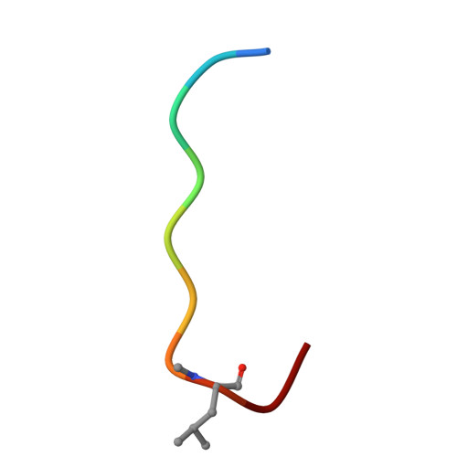
EP67 ligandA [auth D]10Homo sapiensMutation(s): 0 
Entity Groups
Sequence Clusters30% Identity50% Identity70% Identity90% Identity95% Identity100% Identity
Sequence Annotations
Expand
Reference Sequence
Find similar proteins by:|  3D Structure
Entity ID: 2
MoleculeChains  Sequence LengthOrganismDetailsImage
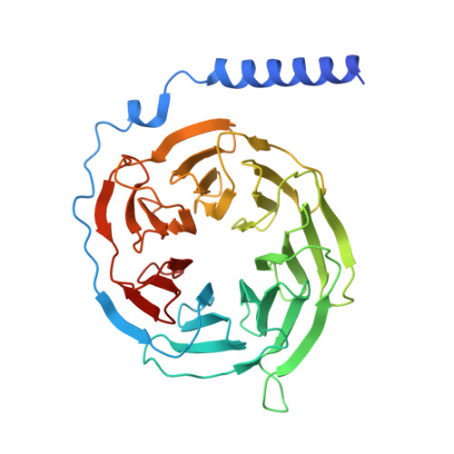
Guanine nucleotide-binding protein G(I)/G(S)/G(T) subunit beta-1350Homo sapiensMutation(s): 0 
Gene Names: GNB1
UniProt & NIH Common Fund Data Resources
Find proteins for P62873 (Homo sapiens)
Explore P62873 
Go to UniProtKB:  P62873
GTEx:  ENSG00000078369 
Entity Groups
Sequence Clusters30% Identity50% Identity70% Identity90% Identity95% Identity100% Identity
UniProt GroupP62873
Sequence Annotations
Expand
Reference Sequence
Find similar proteins by:|  3D Structure
Entity ID: 3
MoleculeChains  Sequence LengthOrganismDetailsImage
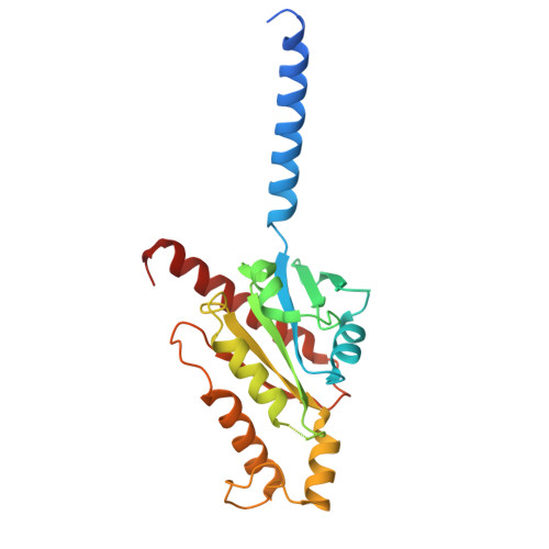
Guanine nucleotide-binding protein G(o) subunit alphaC [auth A]250Homo sapiensMutation(s): 6 
Gene Names: GNAO1
EC: 3.6.5
UniProt & NIH Common Fund Data Resources
Find proteins for P09471 (Homo sapiens)
Explore P09471 
Go to UniProtKB:  P09471
GTEx:  ENSG00000087258 
Entity Groups
Sequence Clusters30% Identity50% Identity70% Identity90% Identity95% Identity100% Identity
UniProt GroupP09471
Sequence Annotations
Expand
Reference Sequence
Find similar proteins by:|  3D Structure
Entity ID: 4
MoleculeChains  Sequence LengthOrganismDetailsImage
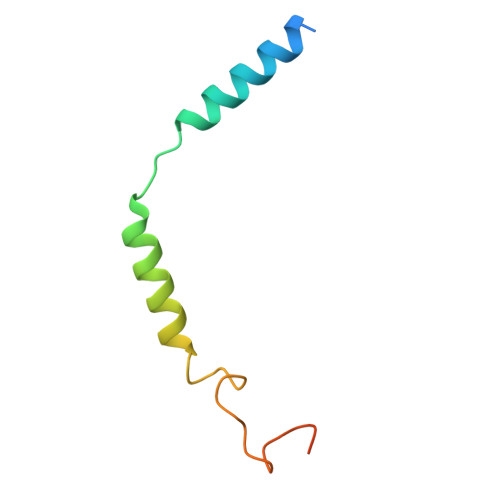
Guanine nucleotide-binding protein G(I)/G(S)/G(O) subunit gamma-2D [auth G]71Homo sapiensMutation(s): 0 
Gene Names: GNG2
UniProt & NIH Common Fund Data Resources
Find proteins for P59768 (Homo sapiens)
Explore P59768 
Go to UniProtKB:  P59768
GTEx:  ENSG00000186469 
Entity Groups
Sequence Clusters30% Identity50% Identity70% Identity90% Identity95% Identity100% Identity
UniProt GroupP59768
Sequence Annotations
Expand
Reference Sequence
Find similar proteins by:|  3D Structure
Entity ID: 5
MoleculeChains  Sequence LengthOrganismDetailsImage
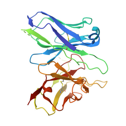
Antibody fragment - ScFv16E [auth H]248Mus musculusMutation(s): 0 
Entity Groups
Sequence Clusters30% Identity50% Identity70% Identity90% Identity95% Identity100% Identity
Sequence Annotations
Expand
Reference Sequence
Find similar proteins by:|  3D Structure
Entity ID: 6
MoleculeChains  Sequence LengthOrganismDetailsImage
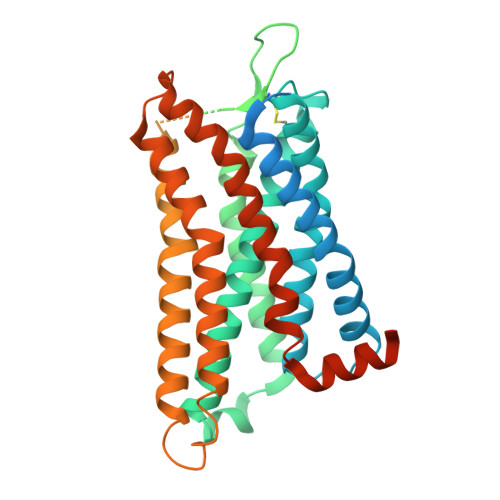
Muscarinic acetylcholine receptor M4,C3a anaphylatoxin chemotactic receptorF [auth C]538Homo sapiensMutation(s): 0 
Gene Names: CHRM4C3AR1AZ3BC3R1HNFAG09
UniProt & NIH Common Fund Data Resources
Find proteins for P08173 (Homo sapiens)
Explore P08173 
Go to UniProtKB:  P08173
GTEx:  ENSG00000180720 
Find proteins for Q16581 (Homo sapiens)
Explore Q16581 
Go to UniProtKB:  Q16581
GTEx:  ENSG00000171860 
Entity Groups
Sequence Clusters30% Identity50% Identity70% Identity90% Identity95% Identity100% Identity
UniProt GroupsP08173Q16581
Sequence Annotations
Expand
Reference Sequence

Small Molecules

Modified Residues  1 Unique
IDChains TypeFormula2D DiagramParent
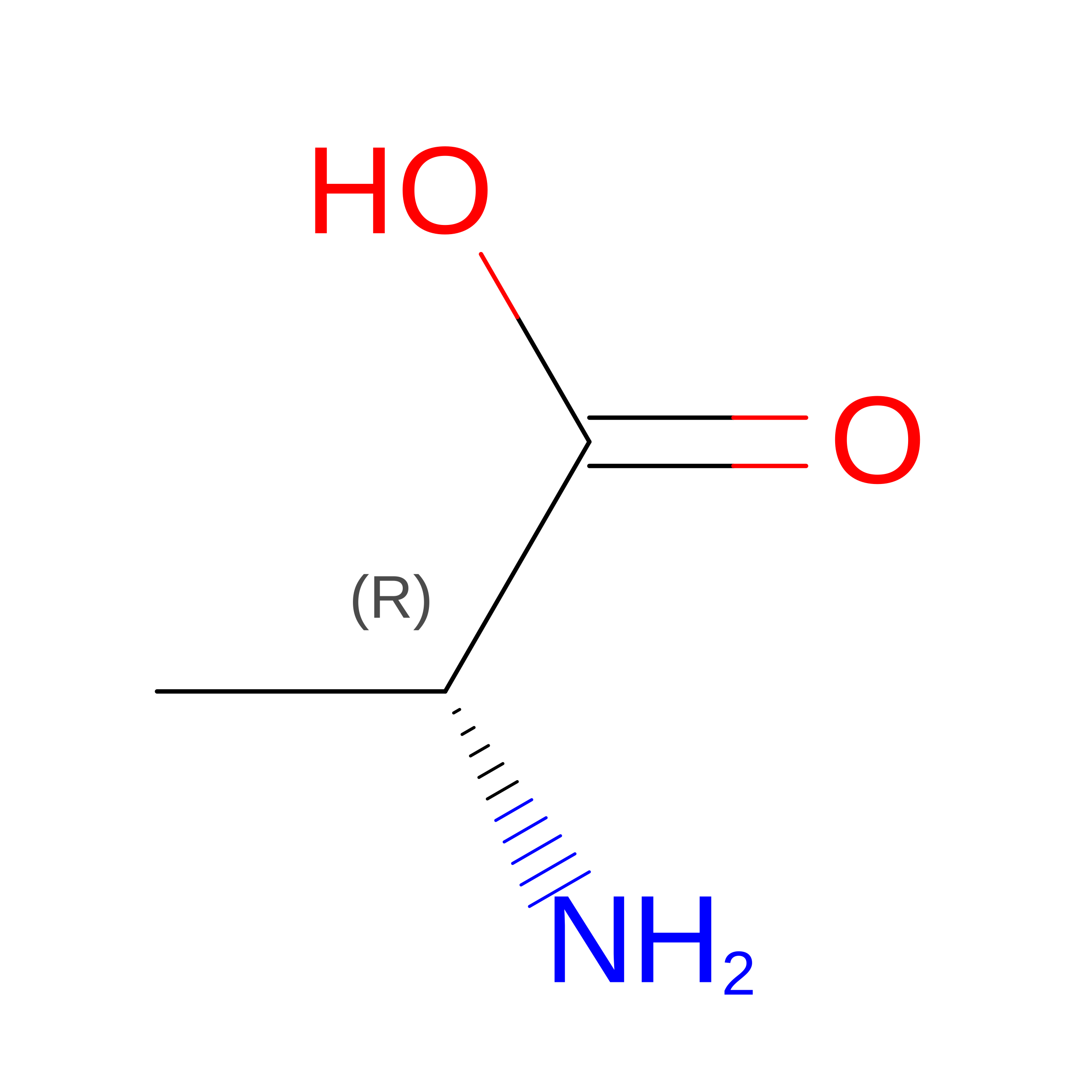
DAL
Query on DAL
A [auth D]D-PEPTIDE LINKINGC3 H7 N O2

--

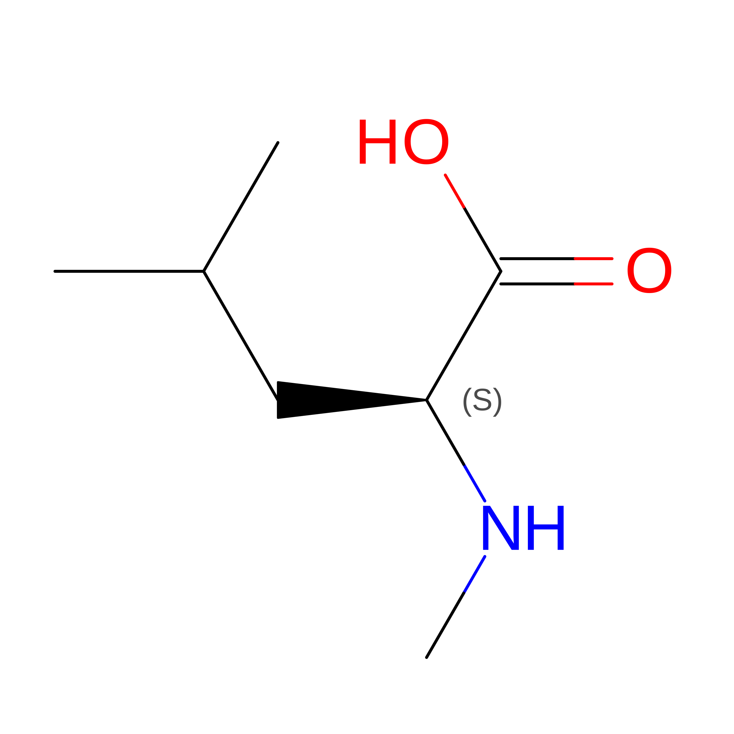
MLE
Query on MLE
A [auth D]L-PEPTIDE LINKINGC7 H15 N O2LEU

Experimental Data & Validation

Experimental Data

  • Method: ELECTRON MICROSCOPY
  • Resolution: 3.43 Å
  • Aggregation State: PARTICLE 
  • Reconstruction Method: SINGLE PARTICLE 
EM Software:
TaskSoftware PackageVersion
MODEL REFINEMENTPHENIX
RECONSTRUCTIONcryoSPARC4.6.2

Structure Validation

View Full Validation Report


Entry History 

& Funding Information

Deposition Data


Funding OrganizationLocationGrant Number
Science and Engineering Research Board (SERB)IndiaIPA/2020/000405
Wellcome TrustUnited KingdomIA/S/20/1/504916
Science and Engineering Research Board (SERB)IndiaCRG/2022/002646

Revision History  (Full details and data files)

  • Version 1.0: 2025-11-26
    Type: Initial release
  • Version 1.1: 2026-07-22
    Changes: Data collection, Database references