9KY5 | pdb_00009ky5

Structure of y+LAT2 bound with Arg


Experimental Data Snapshot

  • Method: ELECTRON MICROSCOPY
  • Resolution: 3.60 Å
  • Aggregation State: PARTICLE 
  • Reconstruction Method: SINGLE PARTICLE 

wwPDB Validation 3D Report Full Report

Validation slider image for 9KY5

This is version 1.1 of the entry. See complete history

Literature

Structural insights into the human system y + L amino acid transporter complex.

Dai, L.Xu, K.Zhang, T.Wang, X.Zeng, Q.Liang, H.Xu, C.Yang, L.Wang, Z.Yan, R.

(2025) Structure 33: 1224-1232.e5

  • DOI: https://doi.org/10.1016/j.str.2025.04.012
  • Primary Citation Related Structures: 
    9KY5, 9LDR

  • PubMed Abstract: 

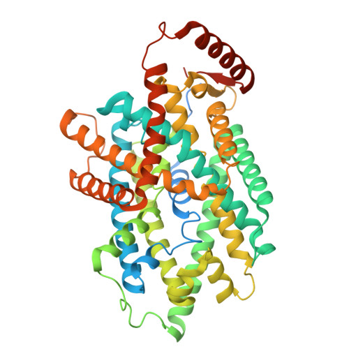
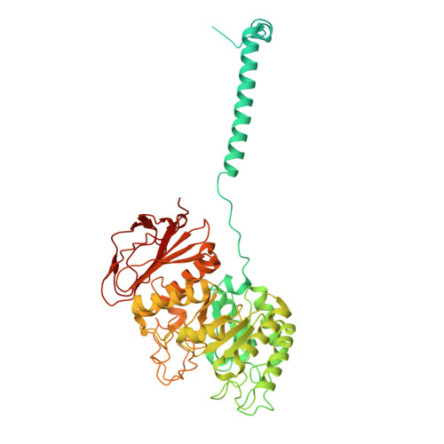
    System y + L facilitates the sodium-independent transport of cationic and sodium-dependent transport of neutral amino acids via heteromeric amino acid transporters. System y + L consists of either SLC7A6 (y + LAT2) or SLC7A7 (y + LAT1) and 4F2hc (SLC3A2). The y + LAT2-4F2hc complex mediates the exchange of L -lysine (Lys), L -arginine (Arg), L -leucine (Leu), and L -glutamine (Gln) and is important for the glutamate-glutamine cycle and ammonia clearance. c-Myc-driven upregulation of y + LAT2 in cancer enhances amino acid uptake and mTORC1 activation, promoting tumor growth. Its transport mechanism has remained unclear. Here, we determined the cryoelectron microscopic (cryo-EM) structures of the y + LAT2-4F2hc complex bound to either Arg or Leu at 3.60 Å and 3.58 Å resolution, respectively, revealing an outward-open conformation. Our structural analysis highlights conformational changes during transport, and functional assays validate critical residues involved in substrate binding and transport. These findings elucidate the molecular mechanism of the system y + L and provide a foundation for developing targeted therapies against y + LAT2.


  • Organizational Affiliation
    • SUSTech Homeostatic Medicine Institute, School of Medicine, Southern University of Science and Technology, Shenzhen, Guangdong Province, China; Key University Laboratory of Metabolism and Health of Guangdong, Institute for Biological Electron Microscopy, Southern University of Science and Technology, Shenzhen, Guangdong Province, China.

Macromolecule Content 

  • Total Structure Weight: 128.95 kDa 
  • Atom Count: 7,277 
  • Modeled Residue Count: 934 
  • Deposited Residue Count: 1,179 
  • Unique protein chains: 2

Macromolecules

Find similar proteins by:|  3D Structure
Entity ID: 1
MoleculeChains  Sequence LengthOrganismDetailsImage
Y+L amino acid transporter 2534Homo sapiensMutation(s): 0 
Gene Names: SLC7A6KIAA0245
UniProt & NIH Common Fund Data Resources
Find proteins for Q92536 (Homo sapiens)
Explore Q92536 
Go to UniProtKB:  Q92536
PHAROS:  Q92536
GTEx:  ENSG00000103064 
Entity Groups
Sequence Clusters30% Identity50% Identity70% Identity90% Identity95% Identity100% Identity
UniProt GroupQ92536
Sequence Annotations
Expand
Reference Sequence
Find similar proteins by:|  3D Structure
Entity ID: 2
MoleculeChains  Sequence LengthOrganismDetailsImage
Amino acid transporter heavy chain SLC3A2645Homo sapiensMutation(s): 0 
Gene Names: SLC3A2MDU1
UniProt & NIH Common Fund Data Resources
Find proteins for P08195 (Homo sapiens)
Explore P08195 
Go to UniProtKB:  P08195
PHAROS:  P08195
GTEx:  ENSG00000168003 
Entity Groups
Sequence Clusters30% Identity50% Identity70% Identity90% Identity95% Identity100% Identity
UniProt GroupP08195
Sequence Annotations
Expand
Reference Sequence

Small Molecules

Ligands 1 Unique
IDChains Name / Formula / InChI Key2D Diagram3D Interactions
ARG

Query on ARG


Download:Ideal Coordinates CCD File
C [auth A]ARGININE
C6 H15 N4 O2
ODKSFYDXXFIFQN-BYPYZUCNSA-O

Experimental Data & Validation

Experimental Data

  • Method: ELECTRON MICROSCOPY
  • Resolution: 3.60 Å
  • Aggregation State: PARTICLE 
  • Reconstruction Method: SINGLE PARTICLE 

Structure Validation

View Full Validation Report


Entry History 

& Funding Information

Deposition Data


Funding OrganizationLocationGrant Number
National Natural Science Foundation of China (NSFC)China--

Revision History  (Full details and data files)

  • Version 1.0: 2025-04-23
    Type: Initial release
  • Version 1.1: 2026-05-13
    Changes: Data collection, Database references