9KPY | pdb_00009kpy

Structure of Phosphopantetheine adenylyltransferase (PPAT) from Enterobacter spp. with the expression tag bound in the substrate binding site of a neighbouring molecule at 2.20 A resolution.


Experimental Data Snapshot

  • Method: X-RAY DIFFRACTION
  • Resolution: 2.20 Å
  • R-Value Free: 
    0.267 (Depositor), 0.286 (DCC) 
  • R-Value Work: 
    0.207 (Depositor) 

Starting Model: experimental
View more details

wwPDB Validation 3D Report Full Report

Validation slider image for 9KPY

Ligand Structure Quality Assessment 


This is version 1.1 of the entry. See complete history

Literature

Protein-tags and their fragments as potent inhibitors of enzymes: Structure of the ternary complex of phosphopantetheine adenylyltransferase from Enterobacter spp. with tag-peptides and phosphonoacetic acid at 2.20 angstrom resolution.

Ahmad, N.Kumar, V.Goel, V.K.Sharma, P.Sharma, S.Singh, T.P.

(2025) Protein Sci 34: e70216-e70216

  • DOI: https://doi.org/10.1002/pro.70216
  • Primary Citation Related Structures: 
    9KPY

  • PubMed Abstract: 

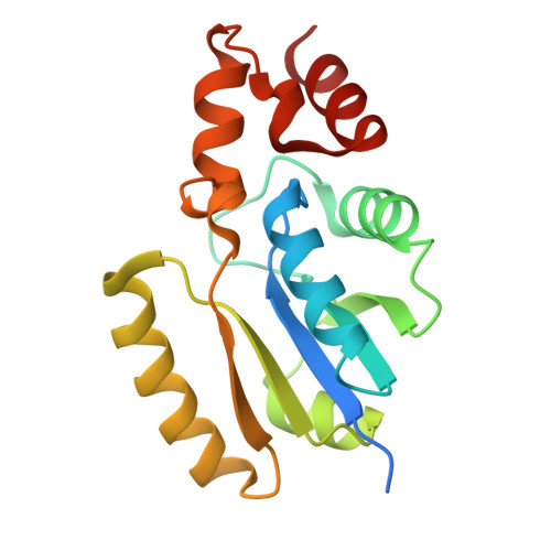
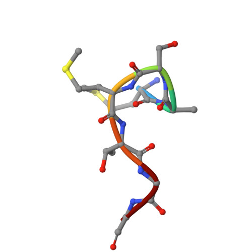
    Phosphopantetheine adenylyltransferase (PPAT) catalyzes the penultimate step of coenzyme A (CoA) biosynthesis pathway by transferring the adenylyl group from adenosine triphosphate (ATP) to 4'-phosphopantetheine (PNS), yielding 3'-dephosphocoenzyme A and pyrophosphate. In this study, the recombinant PPAT from Enterobacter spp. strain 638 (EbPPAT) was purified and co-crystallized with phosphonoacetic acid (PAE). The structure showed the presence of three homodimers AB, CD, and EF in the asymmetric unit. The 14 extra N-terminal residues (Met-14 to Ser-1, 14-mer peptide) from the expression tag were observed in Molecules B and F. These tag-peptides occupied the PNS-binding sites of adjacent Molecules A and E, respectively. Additionally, a heptapeptide (Met-14 to Gly-8) was also observed in the PNS-binding site of Molecule C. Furthermore, two PAE molecules were present in the ATP-binding sites of Molecules B, D, and F, whereas a single PAE molecule was found in Molecules A, C, and E. This showed that tag-peptides blocked the PNS-binding site while PAE blocked the ATP-binding sites. Three peptides of the tag, including 14-mer (Met-14 to Ser-1), heptapeptide (Met-14 to Gly-8) and pentapeptide (Met-14 to Thr-10) were synthesized, and their binding affinities were estimated, which showed the K D values of 5.5 × 10 -5 , 1.8 × 10 -8 , and 7.3 × 10 -9  M, respectively. PAE molecules bound to EbPPAT in the ATP-binding sites with a K D of 4.77 × 10 -4  M. This is the first structure of PPAT with peptides bound in the substrate-binding sites, indicating a novel approach to design peptide inhibitors.


  • Organizational Affiliation
    • Department of Biophysics, All India Institute of Medical Sciences, New Delhi, India.

Macromolecule Content 

  • Total Structure Weight: 117.24 kDa 
  • Atom Count: 8,407 
  • Modeled Residue Count: 989 
  • Deposited Residue Count: 1,045 
  • Unique protein chains: 2

Macromolecules

Find similar proteins by:|  3D Structure
Entity ID: 1
MoleculeChains  Sequence LengthOrganismDetailsImage
Phosphopantetheine adenylyltransferase
A, B, C, D, E
A, B, C, D, E, F
173Enterobacter sp. 638Mutation(s): 0 
Gene Names: coaDEnt638_0105
EC: 2.7.7.3
UniProt
Find proteins for A4W515 (Enterobacter sp. (strain 638))
Explore A4W515 
Go to UniProtKB:  A4W515
Entity Groups
Sequence Clusters30% Identity50% Identity70% Identity90% Identity95% Identity100% Identity
UniProt GroupA4W515
Sequence Annotations
Expand
Reference Sequence
Find similar proteins by:  Sequence   |   3D Structure  
Entity ID: 2
MoleculeChains  Sequence LengthOrganismDetailsImage
HeptapeptideG [auth H]7Enterobacter sp. 638Mutation(s): 0 
Gene Names: coaDEnt638_0105

Small Molecules

Ligands 2 Unique
IDChains Name / Formula / InChI Key2D Diagram3D Interactions
PAE
(Subject of Investigation/LOI)

Query on PAE


Download:Ideal Coordinates CCD File
H [auth A]
J [auth B]
K [auth B]
L [auth C]
M [auth D]
H [auth A],
J [auth B],
K [auth B],
L [auth C],
M [auth D],
N [auth D],
O [auth D],
P [auth E],
Q [auth F],
R [auth F]
PHOSPHONOACETIC ACID
C2 H5 O5 P
XUYJLQHKOGNDPB-UHFFFAOYSA-N
GOL

Query on GOL


Download:Ideal Coordinates CCD File
I [auth B]GLYCEROL
C3 H8 O3
PEDCQBHIVMGVHV-UHFFFAOYSA-N

Experimental Data & Validation

Experimental Data

  • Method: X-RAY DIFFRACTION
  • Resolution: 2.20 Å
  • R-Value Free:  0.267 (Depositor), 0.286 (DCC) 
  • R-Value Work:  0.207 (Depositor) 
Space Group: C 1 2 1
Unit Cell:
Length ( Å )Angle ( ˚ )
a = 137.966α = 90
b = 78.659β = 93.121
c = 107.027γ = 90
Software Package:
Software NamePurpose
REFMACrefinement
MxCuBEdata collection
XDSdata reduction
autoPROCdata scaling
MOLREPphasing
Cootmodel building

Structure Validation

View Full Validation Report


Ligand Structure Quality Assessment 


Entry History 

& Funding Information

Deposition Data


Funding OrganizationLocationGrant Number
Indian Council of Medical ResearchIndiaI-1251

Revision History  (Full details and data files)

  • Version 1.0: 2024-12-11
    Type: Initial release
  • Version 1.1: 2025-08-20
    Changes: Database references