9KL6 | pdb_00009kl6

Crystal structure of NADP-specific glutamate dehydrogenase Gdh1 from Schizosaccharomyces pombe in complex with alpha-iminoglutarate and NADP+


Experimental Data Snapshot

  • Method: X-RAY DIFFRACTION
  • Resolution: 1.45 Å
  • R-Value Free: 
    0.210 (Depositor), 0.213 (DCC) 
  • R-Value Work: 
    0.179 (Depositor), 0.182 (DCC) 
  • R-Value Observed: 
    0.181 (Depositor) 

Starting Model: experimental
View more details

wwPDB Validation 3D Report Full Report

Validation slider image for 9KL6

Ligand Structure Quality Assessment 


This is version 1.2 of the entry. See complete history

Literature

Phosphorylation-mediated regulation of the NADPH-dependent glutamate dehydrogenase, SpGdh1, from Schizosaccharomyces pombe.

Wang, Y.F.Tomita, T.Yoshida, A.Kosono, S.Nishiyama, M.

(2025) J Biological Chem 301: 110422-110422

  • DOI: https://doi.org/10.1016/j.jbc.2025.110422
  • Primary Citation Related Structures: 
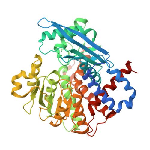
    9KL6

  • PubMed Abstract: 

    Glutamate dehydrogenase from the yeast Schizosaccharomyces pombe (SpGdh1) is a pivotal enzyme that catalyzes the conversion of 2-oxoglutarate and ammonium to glutamate using NADPH as a coenzyme. Although SpGdh1 is phosphorylated at several residues, the impact of phosphorylation on enzyme activity and the underlying molecular mechanisms remains unclear. To elucidate the phosphorylation-mediated regulation of SpGdh1, we determined the crystal structure of SpGdh1 binding 2-iminoglutarate (2-IG) and NADP + . The results of the structural analysis revealed that four serine residues for phosphorylation were located near the active site. Ser252 directly interacted with the 2'-phosphate group of the adenine ribose moiety of NADP + , suggesting that the phosphorylation of Ser252 interfered with NADP + binding. To confirm this hypothesis, we prepared SpGdh1 phosphorylation-mimic (Ser to Glu) variants of SpGdh1 at these four Ser residues. The results of a kinetic analysis revealed that the replacement of these four residues increased the apparent K m NADP(H) value and decreased catalytic efficiency, k cat /K m NADP(H) .In contrast, substitutions decreased the apparent K m NAD(H) value and increased catalytic efficiency, k cat /K m NAD(H) . Therefore, the Ser to Glu replacement caused net shifts in the coenzyme specificities (NADPH to NADH and NADP + to NAD + ) of 55- and 2900-fold, respectively. This is the first study to reveal the effects of the phosphorylation of SpGdh1 on its activity.


  • Organizational Affiliation
    • Graduate School of Agricultural and Life Sciences, The University of Tokyo, Bunkyo-ku, Tokyo, Japan.

Macromolecule Content 

  • Total Structure Weight: 52.32 kDa 
  • Atom Count: 3,937 
  • Modeled Residue Count: 449 
  • Deposited Residue Count: 472 
  • Unique protein chains: 1

Macromolecules

Find similar proteins by:|  3D Structure
Entity ID: 1
MoleculeChains  Sequence LengthOrganismDetailsImage
NADP-specific glutamate dehydrogenase472Schizosaccharomyces pombeMutation(s): 0 
Gene Names: gdh1SPCC622.12c
EC: 1.4.1.4
UniProt
Find proteins for P78804 (Schizosaccharomyces pombe (strain 972 / ATCC 24843))
Explore P78804 
Go to UniProtKB:  P78804
Entity Groups
Sequence Clusters30% Identity50% Identity70% Identity90% Identity95% Identity100% Identity
UniProt GroupP78804
Sequence Annotations
Expand
Reference Sequence

Small Molecules

Ligands 4 Unique
IDChains Name / Formula / InChI Key2D Diagram3D Interactions
NAP
(Subject of Investigation/LOI)

Query on NAP


Download:Ideal Coordinates CCD File
B [auth A]NADP NICOTINAMIDE-ADENINE-DINUCLEOTIDE PHOSPHATE
C21 H28 N7 O17 P3
XJLXINKUBYWONI-NNYOXOHSSA-N
2IT
(Subject of Investigation/LOI)

Query on 2IT


Download:Ideal Coordinates CCD File
D [auth A](2Z)-2-iminopentanedioic acid
C5 H7 N O4
UZWLXPOZNAJCJV-UTCJRWHESA-N
SO4

Query on SO4


Download:Ideal Coordinates CCD File
C [auth A]SULFATE ION
O4 S
QAOWNCQODCNURD-UHFFFAOYSA-L
GOL

Query on GOL


Download:Ideal Coordinates CCD File
E [auth A],
F [auth A]
GLYCEROL
C3 H8 O3
PEDCQBHIVMGVHV-UHFFFAOYSA-N

Experimental Data & Validation

Experimental Data

  • Method: X-RAY DIFFRACTION
  • Resolution: 1.45 Å
  • R-Value Free:  0.210 (Depositor), 0.213 (DCC) 
  • R-Value Work:  0.179 (Depositor), 0.182 (DCC) 
  • R-Value Observed: 0.181 (Depositor) 
Space Group: P 63 2 2
Unit Cell:
Length ( Å )Angle ( ˚ )
a = 96.965α = 90
b = 96.965β = 90
c = 188.258γ = 120
Software Package:
Software NamePurpose
REFMACrefinement
Aimlessdata scaling
XDSdata reduction
PHASERphasing

Structure Validation

View Full Validation Report


Ligand Structure Quality Assessment 


Entry History 

& Funding Information

Deposition Data


Funding OrganizationLocationGrant Number
Japan Society for the Promotion of Science (JSPS)Japan21K05358

Revision History  (Full details and data files)

  • Version 1.0: 2025-07-30
    Type: Initial release
  • Version 1.1: 2025-08-06
    Changes: Database references
  • Version 1.2: 2026-03-04
    Changes: Refinement description