9KGP | pdb_00009kgp

The structure of natural P450BM3-H derived from Bacillus megaterium for catalyzing the steroid DHEA


Experimental Data Snapshot

  • Method: X-RAY DIFFRACTION
  • Resolution: 1.52 Å
  • R-Value Free: 
    0.227 (Depositor), 0.227 (DCC) 
  • R-Value Work: 
    0.221 (Depositor), 0.221 (DCC) 
  • R-Value Observed: 
    0.225 (Depositor) 

wwPDB Validation 3D Report Full Report

Validation slider image for 9KGP

Ligand Structure Quality Assessment 


This is version 1.1 of the entry. See complete history

Literature

Rationally designing P450BM3-H to excavate a novel channel for product exit and enhance overall performance.

Deng, Q.Lu, Z.M.Yuan, Z.Feng, Y.Zhang, L.Shi, J.Xu, Z.Kofas, M.A.G.Li, H.

(2025) Int J Biol Macromol 307: 142162-142162

  • DOI: https://doi.org/10.1016/j.ijbiomac.2025.142162
  • Primary Citation Related Structures: 
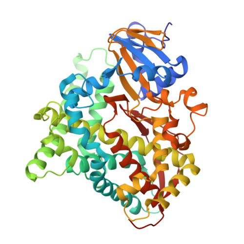
    9KGP

  • PubMed Abstract: 

    P450 dihydroxylase plays a crucial role in steroid drug synthesis by efficiently catalyzing two-step selective hydroxylation reactions. However, natural P450 dihydroxylases are scarce, with poor catalytic performance and intermediate accumulation limiting production. Here, we report a P450 dihydroxylase BM3-H that synthesizes 7α,15α-diOH-DHEA with 76 % selectivity. To enhance 7α,15α-diOH-DHEA synthesis, we engineered a novel exit channel for the intermediate by modifying key residues in the solvent channel. The triple mutant D182K/E143D/V178A exhibited significant improvements in product concentration (10.08-fold), enzymatic activity (2.16-fold), catalytic efficiency (k cat /K m , 42.32-fold), electron transfer rate (k ET , 27.14-fold), and coupling efficiency (CE, 3.93-fold). Molecular dynamics simulations revealed that D182K/E143D/V178A created a novel exit channel for 7α-OH-DHEA, with channel length, polarity, and steric hindrance influencing enzyme performance. Our approach enhances the overall catalytic performance of P450BM3-H by excavating new intermediate product exit channels, providing theoretical guidance for the design of other enzyme molecules.


  • Organizational Affiliation
    • School of Biotechnology, Jiangnan University, Wuxi, People's Republic of China. Electronic address: 7210201002@stu.jiangnan.edu.cn.

Macromolecule Content 

  • Total Structure Weight: 105.39 kDa 
  • Atom Count: 7,928 
  • Modeled Residue Count: 906 
  • Deposited Residue Count: 910 
  • Unique protein chains: 1

Macromolecules

Find similar proteins by:|  3D Structure
Entity ID: 1
MoleculeChains  Sequence LengthOrganismDetailsImage
Bifunctional cytochrome P450/NADPH--P450 reductase
A, B
455Priestia megaterium NBRC 15308 = ATCC 14581Mutation(s): 0 
Gene Names: cyp102A1cyp102BG04_163
EC: 1.14.14.1 (PDB Primary Data), 1.6.2.4 (PDB Primary Data)
UniProt
Find proteins for P14779 (Priestia megaterium (strain ATCC 14581 / DSM 32 / CCUG 1817 / JCM 2506 / NBRC 15308 / NCIMB 9376 / NCTC 10342 / NRRL B-14308 / VKM B-512 / Ford 19))
Explore P14779 
Go to UniProtKB:  P14779
Entity Groups
Sequence Clusters30% Identity50% Identity70% Identity90% Identity95% Identity100% Identity
UniProt GroupP14779
Sequence Annotations
Expand
Reference Sequence

Experimental Data & Validation

Experimental Data

  • Method: X-RAY DIFFRACTION
  • Resolution: 1.52 Å
  • R-Value Free:  0.227 (Depositor), 0.227 (DCC) 
  • R-Value Work:  0.221 (Depositor), 0.221 (DCC) 
  • R-Value Observed: 0.225 (Depositor) 
Space Group: P 1 21 1
Unit Cell:
Length ( Å )Angle ( ˚ )
a = 58.914α = 90
b = 145.713β = 97.513
c = 63.233γ = 90
Software Package:
Software NamePurpose
REFMACrefinement
Aimlessdata scaling
XDSdata reduction
PHASERphasing

Structure Validation

View Full Validation Report


Ligand Structure Quality Assessment 


Entry History 

& Funding Information

Deposition Data


Funding OrganizationLocationGrant Number
National Natural Science Foundation of China (NSFC)China22078126

Revision History  (Full details and data files)

  • Version 1.0: 2025-05-07
    Type: Initial release
  • Version 1.1: 2025-05-21
    Changes: Database references