9JPT | pdb_00009jpt

Local refinement of H14 nanotubes assembled from baculovirus capsid protein


Experimental Data Snapshot

  • Method: ELECTRON MICROSCOPY
  • Resolution: 3.40 Å
  • Aggregation State: HELICAL ARRAY 
  • Reconstruction Method: SINGLE PARTICLE 

wwPDB Validation 3D Report Full Report

Validation slider image for 9JPT

This is version 1.2 of the entry. See complete history

Literature

Structural polymorphism of two-dimensional lattices assembled from baculoviral capsid proteins.

Tian, K.Na, H.Fu, Y.Chong, T.Leng, C.Meng, F.Liang, Y.Wang, M.Hu, Z.Wang, X.Rao, G.Cao, S.

(2025) Virol Sin 40: 935-945

  • DOI: https://doi.org/10.1016/j.virs.2025.11.005
  • Primary Citation Related Structures: 
    9JPR, 9JPS, 9JPT, 9K2O

  • PubMed Abstract: 

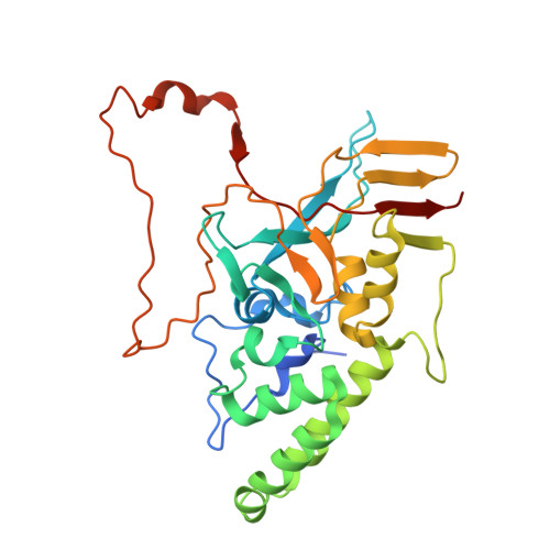
    Protein nanotubes (PNTs) can be regarded as two-dimensional (2D) lattices with p1 or p2 symmetry rolled into tubes. However, attempts to re-assemble their building blocks into stable 2D nanomaterials often fail. Here, starting from two baculoviral capsid proteins, we screened protein variants for the in vitro assembly of various nanotubes and nanosheets. These high-order assemblies were structurally characterized by cryo-electron microscopy techniques. Interfacial analysis of three groups of PNTs revealed that helical heterogeneity is largely the result of the redundancy of p2 symmetry-related contacting interfaces. The assembled nanosheets showed similar interfacial networks to their nanotubular counterparts. In addition, foreign macromolecules could be efficiently displayed on the size-controllable double-layered nanosheets. This study sheds light on the rational design of flexible nanosheets, and it also provides novel 2D protein scaffolds for developing biocompatible materials.


  • Organizational Affiliation
    • State Key Laboratory of Virology and Biosafety, Wuhan Institute of Virology, Chinese Academy of Sciences, Wuhan 430071, China; University of Chinese Academy of Sciences, Beijing 100049, China.

Macromolecule Content 

  • Total Structure Weight: 267.47 kDa 
  • Atom Count: 17,960 
  • Modeled Residue Count: 2,232 
  • Deposited Residue Count: 2,344 
  • Unique protein chains: 1

Macromolecules

Find similar proteins by:|  3D Structure
Entity ID: 1
MoleculeChains  Sequence LengthOrganismDetailsImage
Viral capsid 39 protein
A, B, C, D, E
A, B, C, D, E, F, G, H
293Helicoverpa armigera nucleopolyhedrovirusMutation(s): 0 
UniProt
Find proteins for Q77K63 (Helicoverpa armigera nucleopolyhedrovirus)
Explore Q77K63 
Go to UniProtKB:  Q77K63
Entity Groups
Sequence Clusters30% Identity50% Identity70% Identity90% Identity95% Identity100% Identity
UniProt GroupQ77K63
Sequence Annotations
Expand
Reference Sequence

Experimental Data & Validation

Experimental Data

  • Method: ELECTRON MICROSCOPY
  • Resolution: 3.40 Å
  • Aggregation State: HELICAL ARRAY 
  • Reconstruction Method: SINGLE PARTICLE 
EM Software:
TaskSoftware PackageVersion
MODEL REFINEMENTPHENIX

Structure Validation

View Full Validation Report


Entry History 

& Funding Information

Deposition Data


Funding OrganizationLocationGrant Number
Ministry of Science and Technology (MoST, China)China2022YFC2303300

Revision History  (Full details and data files)

  • Version 1.0: 2025-10-22
    Type: Initial release
  • Version 1.1: 2026-01-21
    Changes: Data collection, Database references
  • Version 1.2: 2026-01-28
    Changes: Data collection, Database references