9JF9 | pdb_00009jf9

Human insulin receptor bound with A62-dimer, Pseudo-arrowhead conformation


Experimental Data Snapshot

  • Method: ELECTRON MICROSCOPY
  • Resolution: 6.26 Å
  • Aggregation State: PARTICLE 
  • Reconstruction Method: SINGLE PARTICLE 

wwPDB Validation 3D Report Full Report

Validation slider image for 9JF9

This is version 1.1 of the entry. See complete history

Literature

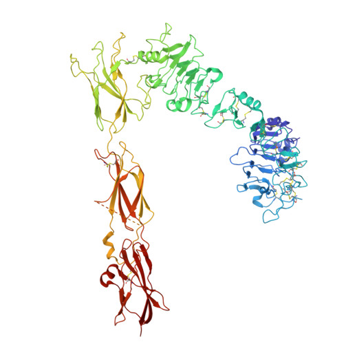
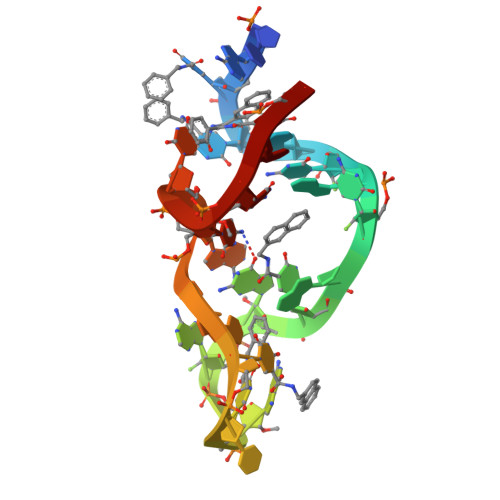
Structural mechanism of insulin receptor activation by a dimeric aptamer agonist.

Kim, J.Na, H.Choi, S.Y.Oh, E.J.Lee, H.Ryu, S.H.Yunn, N.O.Cho, Y.

(2025) Exp Mol Med 57: 1506-1518

  • DOI: https://doi.org/10.1038/s12276-025-01494-1
  • Primary Citation Related Structures: 
    9JF9, 9JFD, 9JHS

  • PubMed Abstract: 

    Insulin binding to the insulin receptor (IR) triggers signaling pathways that regulate glucose uptake and cell growth. In previous work, we identified a DNA aptamer, A62, which partially activates the IR. During engineering aptamers for improved in vivo stability, we discovered that crosslinking two A62 aptamers with linkers of varying lengths led to full phosphorylation of the IR, although activation remained selective to the AKT pathway. Here, to elucidate the mechanism behind this aptamer-induced full activation of the IR, we determined the structure of the IR in complex with a dimeric form of A62 (A62D) linked by an eight-nucleotide connector. We identified three distinct conformations of the IR: arrowhead-shaped, pseudo-arrowhead-shaped and pseudo-gamma-shaped. The pseudo-gamma-shaped conformation closely resembles the structure of a fully active IR bound by a single insulin molecule. In these configurations, only one A62 monomer (A62M) within the A62D dimer binds to the IR dimer. This binding brings the IR monomers into close proximity, promoting intermolecular trans-phosphorylation. Our findings provide valuable structural insights for the development of novel therapeutic strategies targeting the IR.


  • Organizational Affiliation
    • Department of Life Sciences, Pohang University of Science and Technology (POSTECH), Pohang, Republic of Korea.

Macromolecule Content 

  • Total Structure Weight: 226.68 kDa 
  • Atom Count: 14,032 
  • Modeled Residue Count: 1,646 
  • Deposited Residue Count: 1,886 
  • Unique protein chains: 1
  • Unique nucleic acid chains: 1

Macromolecules


Find similar proteins by:|  3D Structure
Entity ID: 1
MoleculeChains  Sequence LengthOrganismDetailsImage
Insulin receptor
A, B
919Homo sapiensMutation(s): 0 
Gene Names: INSR
EC: 2.7.10.1
UniProt & NIH Common Fund Data Resources
Find proteins for P06213 (Homo sapiens)
Explore P06213 
Go to UniProtKB:  P06213
PHAROS:  P06213
GTEx:  ENSG00000171105 
Entity Groups
Sequence Clusters30% Identity50% Identity70% Identity90% Identity95% Identity100% Identity
UniProt GroupP06213
Sequence Annotations
Expand
Reference Sequence
Find similar nucleic acids by:  Sequence
Entity ID: 2
MoleculeChains LengthOrganismImage
Aptamer A62
C, D
24Homo sapiens
Sequence Annotations
Expand
Reference Sequence

Experimental Data & Validation

Experimental Data

  • Method: ELECTRON MICROSCOPY
  • Resolution: 6.26 Å
  • Aggregation State: PARTICLE 
  • Reconstruction Method: SINGLE PARTICLE 
EM Software:
TaskSoftware PackageVersion
MODEL REFINEMENTPHENIX
RECONSTRUCTIONcryoSPARC

Structure Validation

View Full Validation Report


Entry History 

& Funding Information

Deposition Data


Funding OrganizationLocationGrant Number
National Research Foundation (NRF, Korea)Korea, Republic Of--

Revision History  (Full details and data files)

  • Version 1.0: 2025-07-16
    Type: Initial release
  • Version 1.1: 2025-08-20
    Changes: Data collection, Database references