9JDA | pdb_00009jda

taurine transporter


Experimental Data Snapshot

  • Method: ELECTRON MICROSCOPY
  • Resolution: 3.23 Å
  • Aggregation State: PARTICLE 
  • Reconstruction Method: SINGLE PARTICLE 

wwPDB Validation 3D Report Full Report

Validation slider image for 9JDA

This is version 1.0 of the entry. See complete history

Literature

Dimerization and substrate recognition of human taurine transporter.

Zhang, Y.Chen, J.Chen, N.Xiong, H.Zhu, Z.Yang, D.Ge, J.Yu, J.

(2025) Nat Commun 16: 6163-6163

  • DOI: https://doi.org/10.1038/s41467-025-60967-z
  • Primary Citation Related Structures: 
    9JD3, 9JD4, 9JD9, 9JDA, 9JDG, 9JDJ, 9JDL

  • PubMed Abstract: 

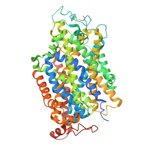
    Taurine is a conditionally essential nutrient and one of the most abundant amino acids in humans, with diverse physiological functions. The cellular uptake of taurine is primarily mediated by the taurine transporter (TauT), and its dysfunction leads to retinal regeneration, cardiomyopathy, neurological and aging-associated disorders. Here we determine structures of TauT in two states: the apo inward-facing open state and the occluded state bound with substrate taurine or γ-aminobutyric acid (GABA). In addition to monomer, the structures also reveal a TauT dimer, where two cholesterol molecules act as "molecular glue", and close contacts of two TM5 from each protomer mediate the dimer interface. In combination with functional characterizations, our results elucidate the detailed mechanisms of substrate recognition, specificity and transport by TauT, providing a structural framework for understanding TauT function and exploring potential therapeutic strategies for taurine-deficiency-related disorders.


  • Organizational Affiliation
    • Interdisciplinary Research Center on Biology and Chemistry, Shanghai Institute of Organic Chemistry, Chinese Academy of Sciences, Shanghai, China.

Macromolecule Content 

  • Total Structure Weight: 70.63 kDa 
  • Atom Count: 4,417 
  • Modeled Residue Count: 552 
  • Deposited Residue Count: 620 
  • Unique protein chains: 1

Macromolecules

Find similar proteins by:|  3D Structure
Entity ID: 1
MoleculeChains  Sequence LengthOrganismDetailsImage
Sodium- and chloride-dependent taurine transporter620Homo sapiensMutation(s): 0 
Gene Names: SLC6A6
Membrane Entity: Yes 
UniProt & NIH Common Fund Data Resources
Find proteins for P31641 (Homo sapiens)
Explore P31641 
Go to UniProtKB:  P31641
PHAROS:  P31641
GTEx:  ENSG00000131389 
Entity Groups
Sequence Clusters30% Identity50% Identity70% Identity90% Identity95% Identity100% Identity
UniProt GroupP31641
Sequence Annotations
Expand
Reference Sequence

Small Molecules

Ligands 6 Unique
IDChains Name / Formula / InChI Key2D Diagram3D Interactions
D12

Query on D12


Download:Ideal Coordinates CCD File
E [auth A]DODECANE
C12 H26
SNRUBQQJIBEYMU-UHFFFAOYSA-N
D10

Query on D10


Download:Ideal Coordinates CCD File
D [auth A]DECANE
C10 H22
DIOQZVSQGTUSAI-UHFFFAOYSA-N
TAU
(Subject of Investigation/LOI)

Query on TAU


Download:Ideal Coordinates CCD File
B [auth A]2-AMINOETHANESULFONIC ACID
C2 H7 N O3 S
XOAAWQZATWQOTB-UHFFFAOYSA-N
OCT

Query on OCT


Download:Ideal Coordinates CCD File
C [auth A],
F [auth A]
N-OCTANE
C8 H18
TVMXDCGIABBOFY-UHFFFAOYSA-N
CL

Query on CL


Download:Ideal Coordinates CCD File
G [auth A]CHLORIDE ION
Cl
VEXZGXHMUGYJMC-UHFFFAOYSA-M
NA

Query on NA


Download:Ideal Coordinates CCD File
H [auth A],
I [auth A]
SODIUM ION
Na
FKNQFGJONOIPTF-UHFFFAOYSA-N

Experimental Data & Validation

Experimental Data

  • Method: ELECTRON MICROSCOPY
  • Resolution: 3.23 Å
  • Aggregation State: PARTICLE 
  • Reconstruction Method: SINGLE PARTICLE 

Structure Validation

View Full Validation Report


Entry History 

& Funding Information

Deposition Data


Funding OrganizationLocationGrant Number
Chinese Academy of SciencesChina--

Revision History  (Full details and data files)

  • Version 1.0: 2025-07-16
    Type: Initial release