9J9K | pdb_00009j9k

Glucosyl transferase NbUGT72AY1 co-crystallized with Scopoletin and UDP2F glucose


Experimental Data Snapshot

  • Method: X-RAY DIFFRACTION
  • Resolution: 3.15 Å
  • R-Value Free: 
    0.258 (Depositor), 0.258 (DCC) 
  • R-Value Work: 
    0.190 (Depositor) 
  • R-Value Observed: 
    0.193 (Depositor) 

Starting Model: experimental
View more details

wwPDB Validation 3D Report Full Report

Validation slider image for 9J9K

Ligand Structure Quality Assessment 


This is version 1.1 of the entry. See complete history

Literature

beta-Carotene alleviates substrate inhibition caused by asymmetric cooperativity.

Liao, J.Shahul Hameed, U.F.Hoffmann, T.D.Kurze, E.Sun, G.Steinchen, W.Nicoli, A.Di Pizio, A.Kuttler, C.Song, C.Catici, D.A.M.Assaad-Gerbert, F.Hoffmann, T.Arold, S.T.Schwab, W.G.

(2025) Nat Commun 16: 3065-3065

  • DOI: https://doi.org/10.1038/s41467-025-58259-7
  • Primary Citation Related Structures: 
    8J2U, 8J2V, 8J2Z, 8J31, 9J9K, 9LRJ

  • PubMed Abstract: 

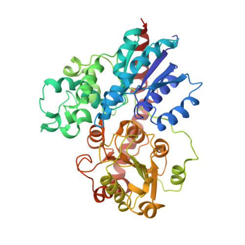
    Enzymes are essential catalysts in biological systems. Substrate inhibition, once dismissed, is now observed in 20% of enzymes 1 and is attributed to the formation of an unproductive enzyme-substrate complex, with no structural evidence of unproductivity provided to date 1-6 . This study uncovers the molecular mechanism of substrate inhibition in tobacco glucosyltransferase NbUGT72AY1, which transfers glucose to phenols for plant protection. The peculiarity that β-carotene strongly attenuates the substrate inhibition of NbUGT72AY1, despite being a competitive inhibitor, allows to determine the conformational changes that occur during substrate binding in both active and substrate-inhibited complexes. Crystallography reveals structurally different ternary enzyme-substrate complexes that do not conform to classical mechanisms. An alternative pathway suggests substrates bind randomly, but the reaction occurs only if a specific order is followed (asymmetric cooperativity). This unreported paradigm explains substrate inhibition and reactivation by competitive inhibitors, opening new research avenues in metabolic regulation and industrial applications.


  • Organizational Affiliation
    • Biotechnology of Natural Products, School of Life Sciences, Technical University of Munich, 85354, Freising, Germany.

Macromolecule Content 

  • Total Structure Weight: 108.1 kDa 
  • Atom Count: 7,493 
  • Modeled Residue Count: 938 
  • Deposited Residue Count: 950 
  • Unique protein chains: 1

Macromolecules

Find similar proteins by:|  3D Structure
Entity ID: 1
MoleculeChains  Sequence LengthOrganismDetailsImage
Glycosyltransferase
A, B
475Nicotiana benthamianaMutation(s): 0 
EC: 2.4.1
UniProt
Find proteins for A0A8K1ZRH3 (Nicotiana benthamiana)
Explore A0A8K1ZRH3 
Go to UniProtKB:  A0A8K1ZRH3
Entity Groups
Sequence Clusters30% Identity50% Identity70% Identity90% Identity95% Identity100% Identity
UniProt GroupA0A8K1ZRH3
Sequence Annotations
Expand
Reference Sequence

Experimental Data & Validation

Experimental Data

  • Method: X-RAY DIFFRACTION
  • Resolution: 3.15 Å
  • R-Value Free:  0.258 (Depositor), 0.258 (DCC) 
  • R-Value Work:  0.190 (Depositor) 
  • R-Value Observed: 0.193 (Depositor) 
Space Group: I 1 2 1
Unit Cell:
Length ( Å )Angle ( ˚ )
a = 101.607α = 90
b = 114.919β = 103.12
c = 104.249γ = 90
Software Package:
Software NamePurpose
REFMACrefinement
XDSdata reduction
XDSdata scaling
PHASERphasing

Structure Validation

View Full Validation Report


Ligand Structure Quality Assessment 


Entry History 

& Funding Information

Deposition Data


Funding OrganizationLocationGrant Number
Other privateSaudi ArabiaKing Abdullah University of Science and Technology

Revision History  (Full details and data files)

  • Version 1.0: 2024-09-04
    Type: Initial release
  • Version 1.1: 2025-04-09
    Changes: Database references, Structure summary